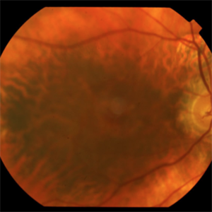
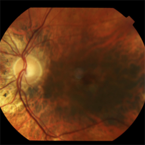
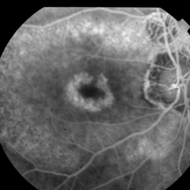
This patient presented complaining of chronically limited central vision. He also had a history of eye trauma OS and had moderately high myopia OS. He had been given a variety of diagnoses in the past. Visual acuities measured 20/40 OD and finger counting OS. Macular exam revealed subtle bulls eye RPE changes that were prominent on fluorescein angiography. Visual field testing showed central scotoma OU. ERG was consistent with cone > rod dysfunction.
-
 Cone-Rod Dystrophy
Aug 15 2015 by Thomas A. Ciulla, MD, MBA, FASRS
Fundus image revealing bull's eye maculopathy.
Photographer: Charlotte Harris
Condition/keywords: cone dystrophy
-
 Cone-Rod Dystrophy
Aug 15 2015 by Thomas A. Ciulla, MD, MBA, FASRS
Fundus image revealing bull's eye maculopathy.
Photographer: Charlotte Harris
Condition/keywords: cone dystrophy
-
 Cone-Rod Dystrophy
Aug 15 2015 by Thomas A. Ciulla, MD, MBA, FASRS
Fundus image revealing bull's eye maculopathy.
Photographer: Charlotte Harris
Condition/keywords: cone dystrophy
-
 Cone-Rod Dystrophy
Aug 15 2015 by Thomas A. Ciulla, MD, MBA, FASRS
Fundus image revealing bull's eye maculopathy.
Photographer: Charlotte Harris
Condition/keywords: cone dystrophy