-
 Macular Degeneration
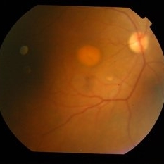
Aug 10 2015 by Mallika Goyal, MD
Fundus of an 82-year-male complaining of distortion of images for 7 years. Bilateral raised circular hypopigmented lesion with smooth edges suggestive of macular dystrophy. Visual acuity was 20/40 each eye. Family history was not significant.
Photographer: Mallika Goyal, MD, Apollo Health City, Hyderabad
Condition/keywords: heredomacular degeneration
-
 Macular Degeneration
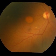
Aug 10 2015 by Mallika Goyal, MD
Fundus of an 82-year-male complaining of distortion of images for 7 years. Bilateral raised circular hypopigmented lesion with smooth edges suggestive of macular dystrophy. Visual acuity was 20/40 each eye. Family history was not significant.
Photographer: Mallika Goyal, MD, Apollo Health City, Hyderabad
Condition/keywords: heredomacular degeneration
-
 Macular Degeneration
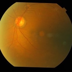
Aug 10 2015 by Mallika Goyal, MD
Fundus of an 82-year-male complaining of distortion of images for 7 years. Bilateral raised circular hypopigmented lesion with smooth edges suggestive of macular dystrophy. Visual acuity was 20/40 each eye. Family history was not significant.
Photographer: Mallika Goyal, MD, Apollo Health City, Hyderabad
Condition/keywords: heredomacular degeneration
-
 Macular Degeneration
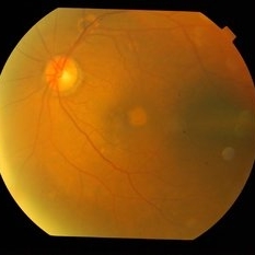
Aug 10 2015 by Mallika Goyal, MD
Fundus of an 82-year-male complaining of distortion of images for 7 years. Bilateral raised circular hypopigmented lesion with smooth edges suggestive of macular dystrophy. Visual acuity was 20/40 each eye. Family history was not significant.
Photographer: Mallika Goyal, MD, Apollo Health City, Hyderabad
Condition/keywords: heredomacular degeneration
A project from the American Society of Retina Specialists