-
 Bilateral Macular Dystrophy
Aug 9 2015 by Mallika Goyal, MD
12-year-old boy presented with left eye vision drop noted for 2 weeks. His mother and maternal grandmother had similar vision problems since young age suggesting possible autosomal dominant inheritance. Both eyes fundus exam revealed macular dystrophy. The left eye macular appearance has changed over the last 3 years becoming more scarred; right eye has remained unchanged. Visual acuity is 20/25 each eye.
Photographer: Mallika Goyal, MD, Apollo Health City, Hyderabad, India
Condition/keywords: heredomacular degeneration
-
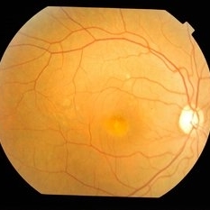
 Bilateral Macular Dystrophy
Aug 9 2015 by Mallika Goyal, MD
12-year-old boy presented with left eye vision drop noted for 2 weeks. His mother and maternal grandmother had similar vision problems since young age suggesting possible autosomal dominant inheritance. Both eyes fundus exam revealed macular dystrophy. The right eye macular appearance has remained unchanged over the 3 years of observation; left eye macular appearance has changed, becoming progressively more scarred. Visual acuity is 20/25 each eye.
Photographer: Mallika Goyal, MD, Apollo Health City, Hyderabad, India
Condition/keywords: heredomacular degeneration
-
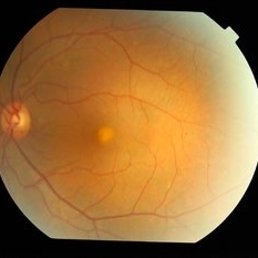
 Bilateral Macular Dystrophy
Aug 9 2015 by Mallika Goyal, MD
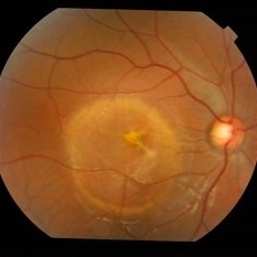
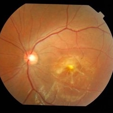
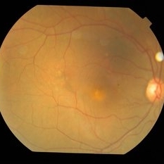
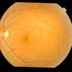
Right eye of a 12-year-old boy at presentation in 2012 shows macular dystrophy. This macula eye appearance did not change on observation for 3 years; left changed over the next 2 years to become more scarred in appearence. Patient's mother and maternal grandmother had similar problems suggesting possible autosomal dominant inheritance.
Photographer: Mallika Goyal, MD, Apollo Health City, Hyderabad
Condition/keywords: heredomacular degeneration
-
 Bilateral Macular Dystrophy
Aug 9 2015 by Mallika Goyal, MD
Right eye of a 12-year-old boy at presentation in 2012 shows macular dystrophy. This macula eye appearance did not change on observation for 3 years; left changed over the next 2 years to become more scarred in appearence. Patient's mother and maternal grandmother had similar problems suggesting possible autosomal dominant inheritance.
Photographer: Mallika Goyal, MD, Apollo Health City, Hyderabad
Condition/keywords: heredomacular degeneration
-
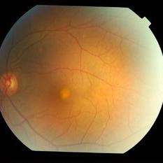
 Bilateral Macular Dystrophy
Aug 9 2015 by Mallika Goyal, MD
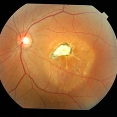
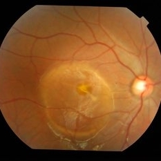
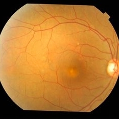
Left eye of a 12-year-old boy at presentation in 2012 shows macular dystrophy. This changed over the next 2 years to become more scarred in appearence. Patient's mother and maternal grandmother had similar problems suggesting possible autosomal dominant inheritance.
Photographer: Mallika Goyal, MD, Apollo Health City, Hyderabad
Condition/keywords: heredomacular degeneration
-
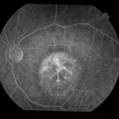
 Bilateral Macular Dystrophy
Aug 9 2015 by Mallika Goyal, MD
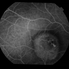
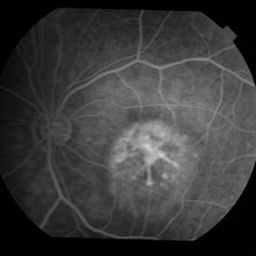
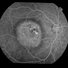
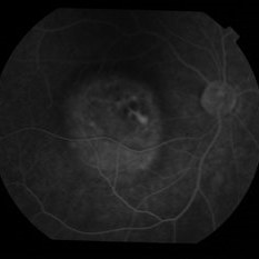
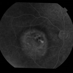
Fluorescein angiogram of the right eye of a 12-year-old boy with bilateral macular dystrophy.
Photographer: Mallika Goyal, MD, Apollo Health City, Hyderabad
Condition/keywords: heredomacular degeneration
-
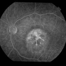
 Bilateral Macular Dystrophy
Aug 9 2015 by Mallika Goyal, MD
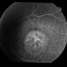
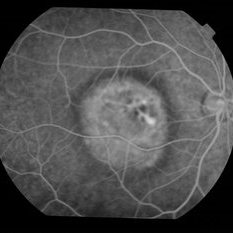
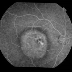
Fluorescein angiogram of the left eye of a 12-year-old boy with bilateral macular dystrophy.
Photographer: Mallika Goyal, MD, Apollo Health City, Hyderabad
Condition/keywords: heredomacular degeneration
-
 Bilateral Macular Dystrophy
Aug 9 2015 by Mallika Goyal, MD
Fluorescein angiogram of the left eye of a 12-year-old boy with bilateral macular dystrophy.
Photographer: Mallika Goyal, MD, Apollo Health City, Hyderabad
Condition/keywords: heredomacular degeneration
-
 Bilateral Macular Dystrophy
Aug 9 2015 by Mallika Goyal, MD
Fluorescein angiogram of the left eye of a 12-year-old boy with bilateral macular dystrophy.
Photographer: Mallika Goyal, MD, Apollo Health City, Hyderabad
Condition/keywords: heredomacular degeneration
-
 Bilateral Macular Dystrophy
Aug 9 2015 by Mallika Goyal, MD
Fluorescein angiogram of the left eye of a 12-year-old boy with bilateral macular dystrophy.
Photographer: Mallika Goyal, MD, Apollo Health City, Hyderabad
Condition/keywords: heredomacular degeneration
-
 Bilateral Macular Dystrophy
Aug 9 2015 by Mallika Goyal, MD
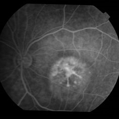
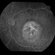
Fluorescein angiogram of the right eye of a 12-year-old boy with bilateral macular dystrophy.
Photographer: Mallika Goyal, MD, Apollo Health City, Hyderabad
Condition/keywords: heredomacular degeneration
-
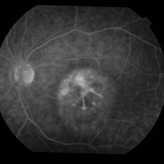
 Bilateral Macular Dystrophy
Aug 9 2015 by Mallika Goyal, MD
Fluorescein angiogram of the right eye of a 12-year-old boy with bilateral macular dystrophy.
Photographer: Mallika Goyal, MD, Apollo Health City, Hyderabad
Condition/keywords: heredomacular degeneration
-
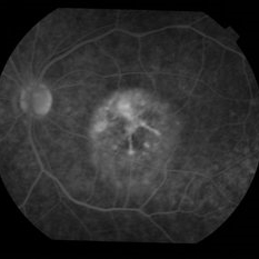
 Bilateral Macular Dystrophy
Aug 9 2015 by Mallika Goyal, MD
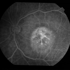
Fluorescein angiogram of the right eye of a 12-year-old boy with bilateral macular dystrophy.
Photographer: Mallika Goyal, MD, Apollo Health City, Hyderabad
Condition/keywords: heredomacular degeneration
-
 Bilateral Macular Dystrophy
Aug 9 2015 by Mallika Goyal, MD
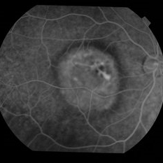
Fluorescein angiogram of the left eye of a 12-year-old boy with bilateral macular dystrophy.
Photographer: Mallika Goyal, MD, Apollo Health City, Hyderabad
Condition/keywords: heredomacular degeneration
-
 Bilateral Macular Dystrophy
Aug 9 2015 by Mallika Goyal, MD
Fluorescein angiogram of the right eye of a 12-year-old boy with bilateral macular dystrophy.
Photographer: Mallika Goyal, MD, Apollo Health City, Hyderabad
Condition/keywords: heredomacular degeneration
-
 Bilateral Macular Dystrophy
Aug 9 2015 by Mallika Goyal, MD
Fluorescein angiogram of the right eye of a 12-year-old boy with bilateral macular dystrophy.
Photographer: Mallika Goyal, MD, Apollo Health City, Hyderabad
Condition/keywords: heredomacular degeneration
-
 Bilateral Macular Dystrophy
Aug 9 2015 by Mallika Goyal, MD
Fluorescein angiogram of the left eye of a 12-year-old boy with bilateral macular dystrophy.
Photographer: Mallika Goyal, MD, Apollo Health City, Hyderabad
Condition/keywords: heredomacular degeneration
-
 Bilateral Macular Dystrophy
Aug 9 2015 by Mallika Goyal, MD
Fluorescein angiogram of the left eye of a 12-year-old boy with bilateral macular dystrophy.
Photographer: Mallika Goyal, MD, Apollo Health City, Hyderabad
Condition/keywords: heredomacular degeneration
-
 Bilateral Macular Dystrophy
Aug 9 2015 by Mallika Goyal, MD
Fluorescein angiogram of the left eye of a 12-year-old boy with bilateral macular dystrophy.
Photographer: Mallika Goyal, MD, Apollo Health City, Hyderabad
Condition/keywords: heredomacular degeneration
-
 Bilateral Macular Dystrophy
Aug 9 2015 by Mallika Goyal, MD
Fluorescein angiogram of the left eye of a 12-year-old boy with bilateral macular dystrophy.
Photographer: Mallika Goyal, MD, Apollo Health City, Hyderabad
Condition/keywords: heredomacular degeneration
-
 Bilateral Macular Dystrophy
Aug 9 2015 by Mallika Goyal, MD
Fluorescein angiogram of the right eye of a 12-year-old boy with bilateral macular dystrophy.
Photographer: Mallika Goyal, MD, Apollo Health City, Hyderabad
Condition/keywords: heredomacular degeneration
-
 Bilateral Macular Dystrophy
Aug 9 2015 by Mallika Goyal, MD
Fluorescein angiogram of the right eye of a 12-year-old boy with bilateral macular dystrophy.
Photographer: Mallika Goyal, MD, Apollo Health City, Hyderabad
Condition/keywords: heredomacular degeneration
-
 Bilateral Macular Dystrophy
Aug 9 2015 by Mallika Goyal, MD
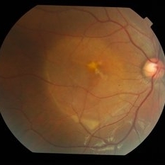
Right fundus of the mother of a 12-year-old boy who presented with bilateral macular dystrophy shows mild macular changes similar to that of her son but less severe than him. Her mother also had visual problems suggesting possible autosomal dominant inheritance.
Photographer: Mallika Goyal, MD, Apollo Health City, Hyderabad
Condition/keywords: heredomacular degeneration
-
 Bilateral Macular Dystrophy
Aug 9 2015 by Mallika Goyal, MD
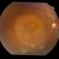
Left fundus of the mother of a 12-year-old boy who presented with bilateral macular dystrophy shows mild macular changes similar to that of her son but less severe than him. Her mother also had visual problems suggesting possible autosomal dominant inheritance.
Photographer: Mallika Goyal, MD, Apollo Health City, Hyderabad
Condition/keywords: heredomacular degeneration
-
 Bilateral Macular Dystrophy
Aug 9 2015 by Mallika Goyal, MD
Left fundus of the mother of a 12-year-old boy who presented with bilateral macular dystrophy shows mild macular changes similar to that of her son but less severe than him. Her mother also had visual problems suggesting possible autosomal dominant inheritance.
Photographer: Mallika Goyal, MD, Apollo Health City, Hyderabad
Condition/keywords: heredomacular degeneration
-
 Bilateral Macular Dystrophy
Aug 20 2015 by Mallika Goyal, MD
OCT of left eye in July 2014, 2 years after presentation. Appearance has altered since presentation; this eye has been treated with 6 injections of avastin and there has been structural and functional improvement.
Photographer: Mallika Goyal, MD
Condition/keywords: macular dystrophy
-
 Bilateral Macular Dystrophy
Aug 20 2015 by Mallika Goyal, MD
OCT of left eye in July 2015, 3 years after presentation. Appearance has altered since presentation; this eye has been treated with 6 injections of avastin and there has been structural and functional improvement.
Photographer: Mallika Goyal, MD
Condition/keywords: macular dystrophy
-
 Bilateral Macular Dystrophy
Aug 20 2015 by Mallika Goyal, MD
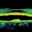
Left eye OCT of a boy with bilateral macular dystrophy at presentation.
Photographer: Mallika Goyal, MD, Apollo Health City, Jubilee Hills, Hyderabad, India
Condition/keywords: macular dystrophy
-
 Bilateral Macular Dystrophy
Aug 20 2015 by Mallika Goyal, MD
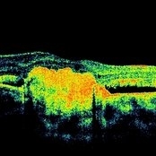
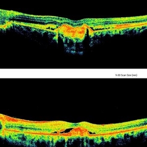
OCT of the left eye of a 12-year-old boy presenting with bilateral macular dystrophy. OCT picture of this eye has changed over 3 years of observation (lower OCT is of 2012, upper one of 2015). This eye was treated with 6 avastin injections over 3 years with resolution of visual symptoms and final VA is 20/25.
Photographer: Mallika Goyal, MD, Apollo Health City, Jubilee Hills, Hyderabad, India
Condition/keywords: macular dystrophy
-
 Bilateral Macular Dystrophy
Aug 20 2015 by Mallika Goyal, MD
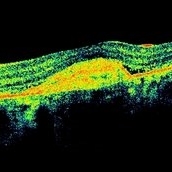
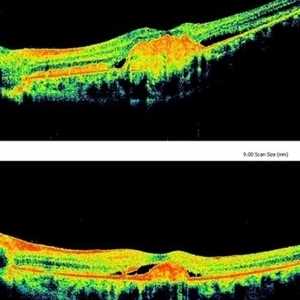
OCT of the left eye of a 12-year-old boy presenting with bilateral macular dystrophy. OCT picture of this eye has changed over the first 2 years of observation (lower OCT is of 2012, upper one of 2014). This eye was treated with 6 Avastin injections over 3 years with resolution of visual symptoms and final VA is 20/25.
Photographer: Mallika Goyal, MD, Apollo Health City, Jubilee Hills, Hyderabad, India
Condition/keywords: macular dystrophy
-
 Bilateral Macular Dystrophy
Aug 20 2015 by Mallika Goyal, MD
OCT of the left eye of a 12-year-old boy presenting with bilateral macular dystrophy. OCT picture of this eye has changed over the last 1 year of management (lower OCT is of 2014, upper one of 2015). This eye was treated with 6 avastin injections over 3 years with resolution of visual symptoms and final VA is 20/25.
Photographer: Mallika Goyal, MD, Apollo Health City, Jubilee Hills, Hyderabad, India
Condition/keywords: macular dystrophy
-
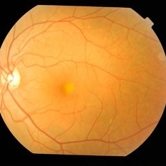
 Bilateral Macular Dystrophy
Aug 20 2015 by Mallika Goyal, MD
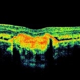
OCT of the right eye of a 12-year-old boy presenting with bilateral macular dystrophy. OCT picture of this eye has remained unchanged over 3 years of observation. VA is 20/25 and there are no visual symptoms.
Photographer: Mallika Goyal, MD, Apollo Health City, Hyderabad
Condition/keywords: macular dystrophy
A project from the American Society of Retina Specialists