-
 Congenital Simple Hamartoma of the RPE Fundus Photo
Aug 3 2015 by Bindu Rajesh
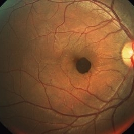
Fundus photograph of a 26-year-old male ,showing a well defined pigmented lesion inferonasal to the fovea suggestive of simple hamartoma.
Imaging device: Visupac
Condition/keywords: congenital, hamartoma, retinal pigment epithelium
-
 Congenital Simple Hamartoma of the RPE Autofluorescence
Aug 3 2015 by Bindu Rajesh
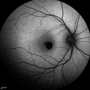
Fundus autofluorescence image of a 26-year-old male depicting hypoautofluorescence nasal to the fovea corresponding to the hamartoma visible clinically.
Imaging device: Heidelberg Spectralis
Condition/keywords: congenital, hamartoma, retinal pigment epithelium
-
 Simple Hamartoma 3D OCT Image
Aug 3 2015 by Bindu Rajesh
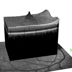
3D OCT image of the hamartoma adjacent to the fovea,showing well defined elevated pigmented lesion with tip protruding into the vitreous.
Imaging device: Heidelberg Spectralis
Condition/keywords: congenital, hamartoma, retinal pigment epithelium
-
 Congenital Simple Hamartoma of RPE
Aug 3 2015 by Bindu Rajesh
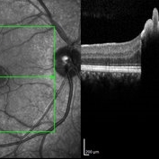
OCT line scan through the hamartoma in a 26-year-old male, showing increased hyperreflectivity in the area of lesion with backshadowing and minimal protrusion into vitreous.
Imaging device: Heidelberg Spectralis
Condition/keywords: congenital, hamartoma, retinal pigment epithelium
A project from the American Society of Retina Specialists