-
 Foveoschisis secondary to high myopia
Mar 13 2015 by Niloofar Piri, MD
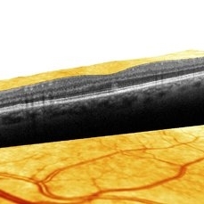
Infrared and HD-OCT of the right eye in a 55-year-old African American female with high myopia (more than -6.00 D), BCVA: 20/25 OU Cartwheel appearance of the fovea in the infrared imaging is visible. HD- OCT demonstartes schisis in different layers of the retina (both NFL and OPL; notice stretching of the Muller cells); VMT is also present . Outer retinal layers are preserved which explains the good vision . She had the same findings in OS.
Photographer: Niloofar Piri, MD
Imaging device: Heidelberg Spectralis
Condition/keywords: high myopia, retinoschisis
-
 AZOOR
AZOOR
Mar 19 2015 by Niloofar Piri, MD
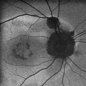
#1: Fundus autofluorescence OD in a patient with AZOOR demonstrates characteristic peripapillary hypoAF as well as concentric rings of hypo and hyper AF in posterior pole .
Imaging device: Heidelberg Spectralis
Condition/keywords: acute zonal occult outer retinopathy (AZOOR)
-
 AZOOR
AZOOR
Mar 19 2015 by Niloofar Piri, MD
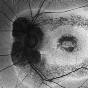
#2 : Fundus autofluorescence OS in the same patient demonstrates more severe changes ; peripapillary hypoAF and concentric rings of hyper and hypo AF in posterior pole
Imaging device: Heidelberg Spectralis
Condition/keywords: acute zonal occult outer retinopathy (AZOOR)
-
 AMN (Acute Macular Neuroretinitis) #2
Apr 28 2019 by Niloofar Piri, MD
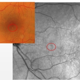
HD-OCT image of 53-year-old man who presented with a superior small paracentral scotoma for 1 month. He had very small hypopigmented area inferior to the fovea and hyporeflectivity on NIR image ( #1). OCT demonstrated a vertical hyper-reflective band extending from OPL to RPE. This form is type 2 AMN which is due to occlusion of deep capillary plexus.
Photographer: Niloofar Piri,MD
Condition/keywords: acute macular neuroretinitis, acute macular neuroretinopathy
-
 AMN (Acute Macular Neurortinitis)
Apr 28 2019 by Niloofar Piri, MD
HD-OCT image of 53-year-old man who presented with a superior small paracentral scotoma for 1 month. He had very small hypopigmented area inferior to the fovea and hyporeflectivity on NIR image ( #1). OCT demonstrated a vertical hyper-reflective band extending from OPL to RPE. This form is type 2 AMN which is due to occlusion of deep capillary plexus. #2
Condition/keywords: acute macular neuroretinitis, acute macular neuroretinopathy
-
 Coats' Disease
Feb 2 2021 by Niloofar Piri, MD
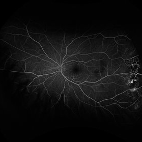
#3 Mid AV phase fluorescein angiography of the same patient demonstrating increasing hyper fluorescence of aneurysmal lesions.
Condition/keywords: Coats' disease, Leber's miliary aneurysm
-
 Coats' Disease
Feb 2 2021 by Niloofar Piri, MD
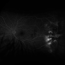
#4 Recirculation phase fluorescein angiography of the same patient demonstrating increased hyperfluorescence and leakage from abnormal vascular lesions in temporal periphery. Note the capillary non perfusion area anteriorly.
Condition/keywords: Coats' disease, Leber's miliary aneurysm
-
 Plaquenil Toxicity
Feb 25 2021 by Niloofar Piri, MD
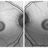
Bilateral fundus autofluorescence of a patient with hydroxycholoroquine (plaquenil) toxicity demonstrating classic bull's eye pattern.
Condition/keywords: bull's eye maculopathy, hydroxychloroquine toxicity, plaquenil toxicity
-
 Plaquenil Toxicity: "Flying Saucer"
Feb 25 2021 by Niloofar Piri, MD
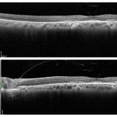
SD- OCT of the same patient with hydroxychloroquine (Plaquenil) toxicity, demonstrating classic "Flying Saucer" pattern secondary to parafoveal RPE atrophy, and outer retinal layer disruption including EZ loss, and outer nuclear layer loss.
Condition/keywords: bull's eye maculopathy, hydroxychloroquine toxicity, plaquenil toxicity
A project from the American Society of Retina Specialists