-
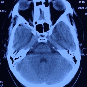 Computed Tomography
Aug 10 2020 by RITESH VERMA
CT scan image section showing fracture of the right greater wing of the sphenoid hampering the blood supply and causing traumatic retinopathy.
Photographer: Dr. Ritesh Verma, Regional institute of Ophthalmology, Rohtak, Haryana, India
Imaging device: CR-2AF CANON
Condition/keywords: CT scan, trauma
-
 Left -Anterior Segment
Aug 10 2020 by RITESH VERMA
Normal anterior segment of the left eye.
Photographer: Dr. Ritesh Verma, Regional institute of Ophthalmology, Rohtak, Haryana, India
Imaging device: CR-2AF CANON
Condition/keywords: anterior segment, normal eye
-
 Left Eye- Posterior Segment
Aug 10 2020 by RITESH VERMA
Normal posterior segment of the left eye.
Photographer: Dr. Ritesh Verma, Regional institute of Ophthalmology, Rohtak, Haryana, India
Imaging device: CR-2AF CANON
Condition/keywords: left eye, normal retina
-
 Right Eye Anterior-Segment
Aug 10 2020 by RITESH VERMA
Anterior segment photograph of right eye showing a silent anterior chamber.
Photographer: Dr. Ritesh Verma, Regional institute of Ophthalmology, Rohtak, Haryana, India
Imaging device: CR-2AF CANON
Condition/keywords: anterior chamber, anterior segment
-
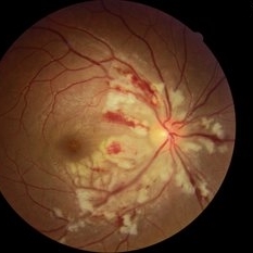 Right Eye-Post Seg
Aug 10 2020 by RITESH VERMA
Fundus photograph of a 27-year-old male with multiple cotton wool spots and hemorrhages in NF layer 6 days post roadside accident.
Photographer: Dr. Ritesh Verma, Regional institute of Ophthalmology, Rohtak, Haryana, India
Imaging device: CR-2AF CANON
Condition/keywords: cotton wool spots, hemorrhage
A project from the American Society of Retina Specialists