-
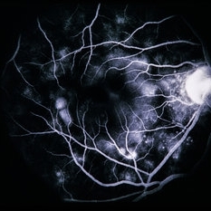
 Vogt-Koyanagi-Harada Disease
Feb 20 2015 by H. Michael Lambert, MD
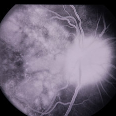
Fluorescein angiogram showing multiple punctate hyperfluorescent dots (starry night) that leak dye into the subretinal space creating multiple serous retinal detachments. There is optic disk leakage.
Condition/keywords: exudative retinal detachment, Vogt-Koyanagi-Harada
-
 Vogt-Koyanagi-Harada Disease
Feb 20 2015 by H. Michael Lambert, MD
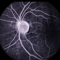
Fluorescein angiogram showing multiple punctate hyperfluorescent dots (starry night) that leak dye into the subretinal space creating multiple serous retinal detachments.
Condition/keywords: exudative retinal detachment, Vogt-Koyanagi-Harada
-
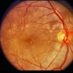
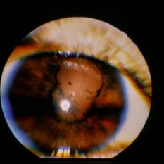
 Vogt-Koyanagi-Harada Disease
Feb 20 2015 by H. Michael Lambert, MD
Color photo showing multifocal detachments of the neurosensory retina with underlying cream colored lesions ( possibly Dalen-Fuchs nodules). Large pocket of subretinal fluid in the macula.
Condition/keywords: exudative detachment, Vogt-Koyanagi-Harada
-
 Vogt-Koyanagi-Harada Disease
Feb 20 2015 by H. Michael Lambert, MD
Color photo showing multifocal detachments of the neurosensory retina with underlying cream colored lesions ( possibly Dalen-Fuchs nodules). Large pocket of subretinal fluid in the macula.
Condition/keywords: exudative retinal detachment, Vogt-Koyanagi-Harada
-
 Vogt-Koyanagi-Harada Disease
Feb 20 2015 by H. Michael Lambert, MD
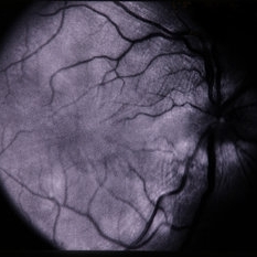
No history. Red free photo showing a normal left eye.
Condition/keywords: exudative retinal detachment, Vogt-Koyanagi-Harada
-
 Vogt-Koyanagi-Harada Disease
Feb 20 2015 by H. Michael Lambert, MD
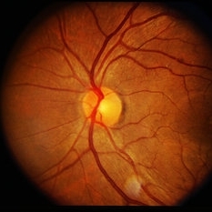
No history. Red free photo showing subretinal fluid in the macular area of the right eye with associated striae of the retina and swelling of the optic nerve.
Condition/keywords: exudative retinal detachment, Vogt-Koyanagi-Harada
-
 Vogt-Koyanagi-Harada Disease
Feb 20 2015 by H. Michael Lambert, MD
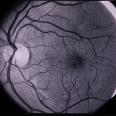
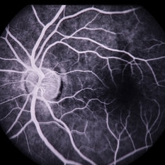
No history. Fluorescein angiogram showing retinal and optic nerve capillary prominence and early leakage.
Condition/keywords: exudative retinal detachment, Vogt-Koyanagi-Harada
-
 Vogt-Koyanagi-Harada Disease
Feb 20 2015 by H. Michael Lambert, MD
No history. Fluorescein angiogram showing retinal and optic nerve capillary prominence and more prominent leakage. Deeper multifocal punctate dots are beginning to show in the macula.
Condition/keywords: exudative retinal detachment, Vogt-Koyanagi-Harada
-
 Vogt-Koyanagi-Harada Disease
Feb 20 2015 by H. Michael Lambert, MD
No history. Fluorescein angiogram showing a normal left eye.
Condition/keywords: exudative retinal detachment, Vogt-Koyanagi-Harada
-
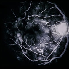
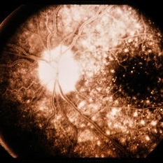
 Vogt-Koyanagi-Harada Disease
Feb 20 2015 by H. Michael Lambert, MD
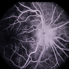
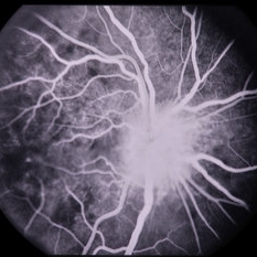
Fluorescein angiogram showing multiple punctate hyperfluorescent dots (starry night) that leak dye into the subretinal space creating multiple serous retinal detachments, becoming confluent in the macular area of the right eye. The optic nerve shows profuse leakage.
Condition/keywords: exudative retinal detachment, Vogt-Koyanagi-Harada
-
 Vogt-Koyanagi-Harada Disease
Feb 20 2015 by H. Michael Lambert, MD
No history. Fluorescein angiogram showing a normal left eye.
Condition/keywords: exudative retinal detachment, Vogt-Koyanagi-Harada
-
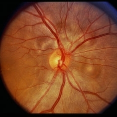
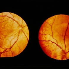
 Vogt-Koyanagi-Harada Disease
Feb 20 2015 by H. Michael Lambert, MD
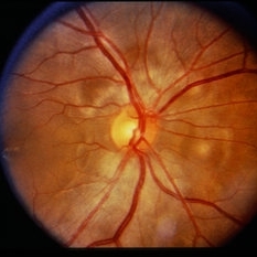
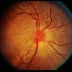
No history. Color photo showing a swollen optic nerve and a large collection of subretinal fluid in the macular area and around the optic nerve of the right eye.
Condition/keywords: exudative retinal detachment, Vogt-Koyanagi-Harada
-
 Vogt-Koyanagi-Harada Disease
Feb 20 2015 by H. Michael Lambert, MD
No history. Color photo showing a normal left eye.
Condition/keywords: exudative retinal detachment, Vogt-Koyanagi-Harada
-
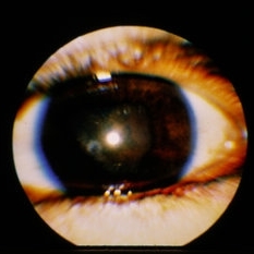
 Vogt-Koyanagi-Harada Disease
Feb 20 2015 by H. Michael Lambert, MD
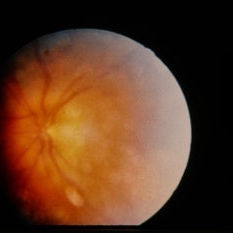
29-year-old female. 20/20 and 20/200. Recurrent uveitis. Color photo showing poor visualization and swelling of the left optic nerve.
Condition/keywords: exudative retinal detachment, Vogt-Koyanagi-Harada
-
 Vogt-Koyanagi-Harada Disease
Feb 20 2015 by H. Michael Lambert, MD
29-year-old female. 20/20 and 20/200. Recurrent uveitis.
Condition/keywords: Vogt-Koyanagi-Harada
-
 Vogt-Koyanagi-Harada Disease
Feb 20 2015 by H. Michael Lambert, MD
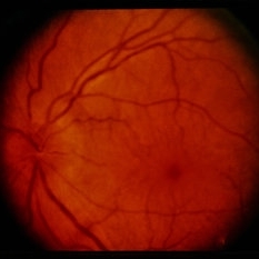
No history. Color photo showing multiple retinal detachments throughout the posterior right eye with prominent retinal striae. There is possible swelling of the optic nerve. The optic nerve may be swollen.
Condition/keywords: exudative retinal detachment, Vogt-Koyanagi-Harada
-
 Vogt-Koyanagi-Harada Disease
Feb 20 2015 by H. Michael Lambert, MD
Dalen-Fuchs' nodule. A granuloma made up of epithelioid cells and lymphocytes between Bruch’s membrane and the retinal pigment epithelium.
Condition/keywords: exudative retinal detachment, Vogt-Koyanagi-Harada
-
 Vogt-Koyanagi-Harada Disease
Feb 20 2015 by H. Michael Lambert, MD
29-year-old female. 20/20 and 20/200. Recurrent uveitis.
Condition/keywords: exudative retinal detachment, Vogt-Koyanagi-Harada
A project from the American Society of Retina Specialists