-
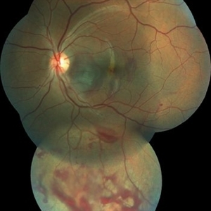
 24 Hours Post Scleral Wound Closure+ Scleral Buckle+25 g Vitrectomy+Silicon Oil
Jan 23 2015 by Carlos Quezada-Ruiz, MD, FASRS
24 hours post op fundus photograph of a 43-year-old man who had perforating injury to the right eye with a small piece of plastic while he was hammering. OD LP, subconjunctival hemorrhage, clear cornea, hyphema, irido and ciclodyalisis as well as a luxated lens with traumatic cataract and a dense vitreous hemorrhage. B-US showed rhegmatogenous retinal detachment with a tear and a big inferior hemorrhagic choroidal detachment. 360 peritomy revealed 2-entry scleral wounds were found in zone II (M V and M VI) and closure was performed. 25 G PPV was performed with the infusion canal placed in the AC through the limbus. Lensectomy and removal of a dense recent vitreous hemorrhage revealed a white detached retina with an exit wound through the temporal inferior segment of the optic nerve with a nasal GRT and sub retinal hemorrhage as well as temporal inferior choroidal, PVD was induced and PFOs helped stabilizing the retina while vitrectomy and sub-retinal hemorrhage was removed through the GRT. Fluid air exchange was made and 360 endolaser over the buckle indentation was done and silicon oil was used as endotamponade. This picture was taken 24 hrs after the surgery.
Photographer: Lilibeth Rodriguez, Instituto de la Visión. Torreon, Mexico.
Condition/keywords: central retinal artery occlusion (CRAO), giant retinal tear, trauma
-
 Choroidal Rupture After Blunt Trauma
Feb 3 2015 by Carlos Quezada-Ruiz, MD, FASRS
OS fundus photograph reconstruction of a 17-year-old man who received "a punch or two" to the face during a street fight.
Photographer: Valeria, Instituto de la Visión. Torreon, Mexico.
Condition/keywords: blunt trauma, choroidal rupture
-
 Ischemic CRVO
Jul 28 2018 by Carlos Quezada-Ruiz, MD, FASRS
Fundus photograph of a 75-year-old woman with hypertension and dyslipidemia who presented to the clinic with sudden decrease in vision on her right eye.
Condition/keywords: central retinal vein occlusion (CRVO)
A project from the American Society of Retina Specialists