-
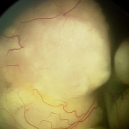
 Bacterial endophthalmitis
Jun 23 2020 by Thirumalesh Mochi Basavaraj, MD
Intraoperative pictures showing bacterial colonies, lab confirmed gram positive cocci(inset).
Imaging device: Leica microsurgery microscope
Condition/keywords: endophthalmitis, intraoperative
-
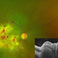
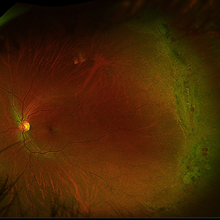
 Coats' Disease Stage 2A
Jun 25 2020 by Thirumalesh Mochi Basavaraj, MD
Fundus photograph (montage) of 9-year-old child with macular exudation. Telangiectic vessels seen. Please note saccular and beaded aneurysmal dilatation of vessels temporally.
Photographer: Puttaswamy
Imaging device: DRI OCT Triton SSOCT- Topcon
Condition/keywords: Coats' disease, idiopathic macular telangiectasia, macular exudates
-
 Crystalline Retinopathy
Jun 27 2020 by Thirumalesh Mochi Basavaraj, MD
23-year-old asymptomatic female, came for routine examination. Inset shows OCT image with hyperreflective lesion scattered throughout the retina, choroidal sclerosis can also be noted.
Photographer: Ravikrishna, Puttaswamy
Imaging device: Heidelberg Spectralis
Condition/keywords: crystalline retinopathy
-
 Dry Age Related Macular Degeneration
Jun 27 2020 by Thirumalesh Mochi Basavaraj, MD
Multiple drusens seen at macula, OCT shows sub RPE deposits of hyper-reflective material.
Photographer: Ravikrishna, Puttaswamy
Imaging device: Heidelberg Spectralis
Condition/keywords: age-related macular degeneration (AMD)
-
 Solar Retinopathy
Jun 27 2020 by Thirumalesh Mochi Basavaraj, MD
Fundus photo showing eclipse burn, Inset OCT picture shows loss of photoreceptor layer.
Photographer: Ravikrishna, Puttaswamy
Imaging device: Heidelberg Spectralis
Condition/keywords: solar retinopathy
-
 Proliferative Diabetic Retinopathy Showing NVD
Aug 12 2020 by Thirumalesh Mochi Basavaraj, MD
Fundus photo showing neovascularisation of disc with OCTA (full disc). Inset shows segmentation level.
Photographer: Ravikrishna, Puttaswamy
Imaging device: DRI OCT triton SSOCT-Topcon
Condition/keywords: neovascularization of the disc (NVD), proliferative diabetic retinopathy (PDR)
-
 Diagnosed Case of Sjogren Larsson
Aug 12 2020 by Thirumalesh Mochi Basavaraj, MD
Hyperreflective deposits seen in inner plexiform layers. Sjogren Larsson is inborn error of lipid metabolism caused by mutation in FADH gene which leads to MCFA, LCFA build up specifically in the membranes of skin and brain. This picture shows shows deposits in IPL.
Photographer: Ravikrishna, Puttaswamy
Imaging device: DRI OCT triton SSOCT-Topcon
Condition/keywords: Sjogren's syndrome
-
 OCTA View of Florid Neovascularization and Enface View
Aug 20 2020 by Thirumalesh Mochi Basavaraj, MD
Image shows florid neovascularization of disc on OCTA. En face imaging shows florid neovascularization along with preretinal bleed.
Photographer: Puttaswamy
Imaging device: DRI OCT Triton SSOCT- Topcon
Condition/keywords: neovascularization of the disc (NVD)
-
 Retinoblastoma
Jun 9 2021 by Thirumalesh Mochi Basavaraj, MD
Large retinoblastoma.
Photographer: Puttaswamy ,Narayana Nethralaya
Imaging device: Retcam shuttle
Condition/keywords: retinoblastoma
-
 Pupillary View of Retinoblastoma
Jun 9 2021 by Thirumalesh Mochi Basavaraj, MD
Group D retinoblastoma.
Photographer: Puttaswamy, Narayana Nethralaya
Imaging device: Retcam Shuttle
Condition/keywords: retinoblastoma
-
 Multifocal Retinoblastoma
Jun 9 2021 by Thirumalesh Mochi Basavaraj, MD
3-year-old kid with multifocal retinoblastoma.
Photographer: Puttaswamy , Narayana Nethralaya , Bangalore
Imaging device: Retcam
Condition/keywords: retinoblastoma
-
 Multifocal Retinoblastoma
Jun 9 2021 by Thirumalesh Mochi Basavaraj, MD
Inferotemporal tumor is blanched immediately post TTT, in comparison to the nasal tumor which has not received TTT.
Photographer: Puttaswamy, Narayana Nethralaya, Bangalore
Imaging device: Retcam
Condition/keywords: retinoblastoma, tumor
-
 Retinoblastoma with Exudative Retinal Detachment
Jun 9 2021 by Thirumalesh Mochi Basavaraj, MD
A case of retinoblastoma with exudative retinal detachment and subretinal seeding.
Photographer: Puttaswamy, Narayan Nethralaya, Bangalore
Imaging device: Retcam
Condition/keywords: exudative retinal detachment
-
 Macular Retinoblastoma
Jun 9 2021 by Thirumalesh Mochi Basavaraj, MD
Macular retinoblastoma.
Photographer: Puttaswamy, Narayana Nethralaya
Imaging device: Retcam
Condition/keywords: tumor
-
 Vitreous Seeds
Jun 9 2021 by Thirumalesh Mochi Basavaraj, MD
Vitreous seeds in a case of retinoblastoma.
Photographer: Puttaswamy, Narayana Nethralaya, Bangalore
Imaging device: retcam
Condition/keywords: retinoblastoma
-
 Cilioretinal Artery Occlusion
Jun 9 2021 by Thirumalesh Mochi Basavaraj, MD
Fundus photograph of 43-year-old women with a Y-shaped clot occluding the trunk and branches of the cilioretinal artery.
Photographer: Puttuswamy , Narayana Nethralaya , Bangalore
Condition/keywords: cilioretinal artery occlusion
-
 Cilioretinal Artery Sparing CRAO
Jun 9 2021 by Thirumalesh Mochi Basavaraj, MD
Cilio retinal artery sparing CRAO in a 40-year-old gentleman post surgery for varicose veins.
Photographer: Puttuswamy
Condition/keywords: central retinal artery occlusion (CRAO)
-
 Choroidal Osteoma
Jan 3 2022 by Thirumalesh Mochi Basavaraj, MD
Fundus photograph of a young female in her second decade with a choroidal mass lesion with calcification suggestive of choroidal osteomalacia.
Photographer: Putta Swamy, Narayana Nethralaya
Imaging device: Topcon DRI Triton
Condition/keywords: macular choroidal osteoma
-
 Macular Hole
Jan 3 2022 by Thirumalesh Mochi Basavaraj, MD
Swept source OCT of a 65-year-old patient with posterior hyaloid separation and a large macular hole with undermined edges.
Photographer: Puttaswamy Narayana Nethralaya Bangalore
Imaging device: Topcon Dri Triton
Condition/keywords: large macular hole, posterior cyclitis hyaloid separation, swept source OCT
-
 Large Macular Hole: Pre-op and Post-op
Jan 4 2022 by Thirumalesh Mochi Basavaraj, MD
Swept source OCT of a 65-yea-old patient with a large macular hole before and after surgery.
Photographer: Puttaswamy Narayana Nethralaya
Imaging device: Topcon Dri Triton
Condition/keywords: macular hole
-
Thirumalesh Mochi Basavraj - Thirumalesh MB.mp4
Jan 10 2022 by Thirumalesh Mochi Basavaraj, MD
Removal of fibrovascular membrane with a bimanual dissection maneuver is demonstrated to release traction in a patient with proliferative diabetic retinopathy with tractional retinal detachment.
Photographer: Thirumalesh Mochi Basavaraj
Condition/keywords: bimanual dissection, fibrovascular membrane, Retinal Detachment, tractional retinal detachment
-
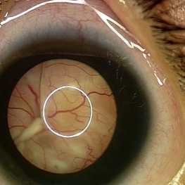
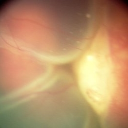
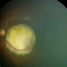
 Capillary Heamngioma of the Optic Disc
Jun 30 2022 by Thirumalesh Mochi Basavaraj, MD
fundus image showing a large Capillary Hemangioma at the Optic Disc
Photographer: Thirumalesh Mochi Basavaraj
Imaging device: intraoperative picture
Condition/keywords: Hemangioma, Von Hippel-Lindau
-
 Choroidal Granuloma
Jul 17 2023 by Thirumalesh Mochi Basavaraj, MD
Wide filed image of a tubercular choroidal granuloma
Photographer: Puttaswamy N K , Narayana Nethralaya, Bangalore
Condition/keywords: choroidal tuberculoma, tubercular choroidal granuloma
-
 Tubercular Granuloma/Choroidal Granuloma
Jul 17 2023 by Thirumalesh Mochi Basavaraj, MD
Composite Optos+SSOCT image Tubercular granuloma
Photographer: Puttaswamy N K ,Narayana Nethralaya, Bangalore
Condition/keywords: composite image OCT and widefiled image, tubercular choroidal granuloma
-
 Retinitis Pigmentosa
Aug 18 2023 by Thirumalesh Mochi Basavaraj, MD
Fundus image of a 30 Year-old young man with night blindness showing a waxy pale disc, attenuated arterioles and mid peripheral pigmentary clumps arranged like bony spicules
Photographer: Puttaswamy
Condition/keywords: bone spicule, Retinitis Pigmentosa, RPE65
-
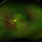
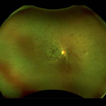
 Albinism
Feb 6 2024 by Thirumalesh Mochi Basavaraj, MD
12 year old child with ocular albinism showing the underlying choroidal vasculature.
Photographer: Puttaswamy
Condition/keywords: ocular albinism
-
 Choroidal Detachment
Feb 6 2024 by Thirumalesh Mochi Basavaraj, MD
60 year old gentleman post trab, presenting with serous choroidal detachment.
Photographer: Puttaswamy
Condition/keywords: serous choroidal detachment
-
 Journey of PDR
Feb 6 2024 by Thirumalesh Mochi Basavaraj, MD
32 year old type 1 diabetic patient with florid neovascularisation with vitreous and subhyaloid hemorrhage, after laser and after mivs surgery.
Photographer: Puttaswamy
Condition/keywords: Diabetic Tractional Retinal Detachment involving the Macula
-
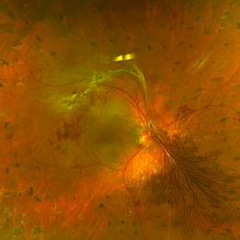
 Florid-NVD
Feb 6 2024 by Thirumalesh Mochi Basavaraj, MD
Young Type 1 diabetic with florid neovascularisation at the optic disc.
Photographer: Puttaswamy
Condition/keywords: NEOVASCULARISATION OF DISC, proliferative diabetic retinopathy (PDR)
-
 Proliferative Diabetic Retinopathy with HRC Lasered
Feb 6 2024 by Thirumalesh Mochi Basavaraj, MD
60 Year Old Type 2 Diabetic with Florid Neovascularisation at the Disc.
Photographer: Puttaswamy
Condition/keywords: florid type PDR, Neovascularisation at the Disc (NVD)
-
 Tractional Retinal Detachment
Feb 6 2024 by Thirumalesh Mochi Basavaraj, MD
22 year old Type 1 DM with a Macular Tractional Retinal Detachment.
Photographer: Puttaswamy
Condition/keywords: Diabetic Tractional Retinal Detachment involving the Macula
-
 Von Hippel-Lindau
Feb 6 2024 by Thirumalesh Mochi Basavaraj, MD
8 Year old child with Multiple Capillary Haemangiomas with Exudative retinal detachment.
Photographer: Puttaswamy
Condition/keywords: exudative detachment, Von Hippel-Lindau
-
 Lattice With Holes
Feb 6 2024 by Thirumalesh Mochi Basavaraj, MD
25 year old myopic patient with extensive lattice degeneration with multiple atrophic holes.
Photographer: Puttaswamy
Condition/keywords: atrophic retinal hole, High Myopia, peripheral lattice degeneration
-
 Lattice With Hole
Feb 6 2024 by Thirumalesh Mochi Basavaraj, MD
25 year old myopic patient with extensive lattice degeneration with multiple atrophic holes.
Photographer: Puttaswamy
Condition/keywords: atrophic hole, High myopia, lattice degeneration
-
 Giant Retinal Tear with Bare Choroid
Dec 13 2024 by Thirumalesh Mochi Basavaraj, MD
Intra-operative view of a Pediatric Giant Retinal Tear with a view of the Bare Choroid Superiorly.
Photographer: Thirumalesh Mochi Basavaraj
Imaging device: Leica Proveo 8
Condition/keywords: GIANT RETINAL TEAR, Retinal Detachment
-
 Massive RIOFB
Dec 16 2024 by Thirumalesh Mochi Basavaraj, MD
A large 23 mm intraocular foreign body post removal
Photographer: Dr Thirumalesh Mochi Basavaraj
Imaging device: iPhone 12
Condition/keywords: Massive 23 mm Iron FB
-
 RIOFB Ferrous
Dec 17 2024 by Thirumalesh Mochi Basavaraj, MD
Radiographic images AP, lateral view, and 3D CT reconstruction along with the extracted 23 mm Iron foreign body from a 28 year old patient who was injured while working in a stone quarry.
Photographer: Thirumalesh Mochi Basavaraj
Condition/keywords: Ferrous FB, RIFOFB
-
 Large Imapacted Ferrous Intraocular Foriegn Body
Dec 17 2024 by Thirumalesh Mochi Basavaraj, MD
Composite image consisting of Radiographic images AP, lateral view, and 3D CT reconstruction , intra operative image ,along with the extracted 23 mm Iron foreign body from a 28 year old patient who was injured while working in a stone quarry.
Photographer: Thirumalesh Mochi Basavaraj
Condition/keywords: Metallic RIOFB
-
 Coats` Disease
Dec 18 2024 by Thirumalesh Mochi Basavaraj, MD
Fundus photo graph of a 6 year old child with exudative retinal detachment with sub retinal lipid exudation and peripheral telengectasia.
Photographer: Puttaswamy N K
Condition/keywords: exudative detachment, peripheral telangiectasia
-
 Sub-Retinal Blood Air and TPA
Jan 31 2025 by Thirumalesh Mochi Basavaraj, MD
Intra Operative View of a 76 year old gentleman with Submacular bleed treated with Sub Retinal TPA, Ranibizumab and air, one can appreciate at multiple levels
Photographer: Thirumalesh Mochi Basavaraj
Condition/keywords: submacular hemorrhage, tissue plasminogen activator (tPA)
-
 Posterior Vitreous Detachment
Jan 31 2025 by Thirumalesh Mochi Basavaraj, MD
Intraoperative view of Triamcinolone-assisted posterior vitreous detachment.
Photographer: Thirumalesh Mochi Basavaraj
Condition/keywords: PVD induction, triamcinolone
-
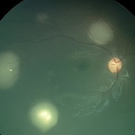
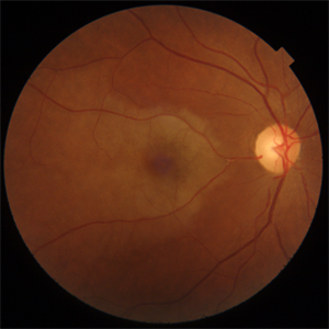
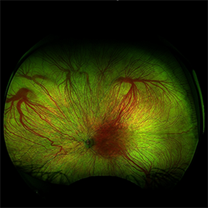
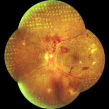
 Eales Disease
Jan 31 2025 by Thirumalesh Mochi Basavaraj, MD
Ultra-wide field image of a 24 year old young healthy adult male with a visible sea fan neovascularization with partial PVD with vitreous and subhyaloid hemorrhage.
Photographer: Puttaswamy
Condition/keywords: Eales disease, sea fan, Ultra-wide field retinal imaging
-
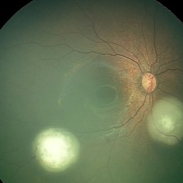
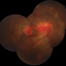
 Eales Disease
Jan 31 2025 by Thirumalesh Mochi Basavaraj, MD
Ultra wide field image of a 24 year-old young healthy adult male with a visible sea fan neovascularization with partial PVD secondary to Scatter LASER photocoagulation with Vitreous and subhyaloid hemorrhage.
Photographer: Puttaswamy N K
Condition/keywords: Eales disease, Neovascularisation elsewhere (NVE), sea fan
-
 Roth Spots Everywhere
Apr 23 2025 by Thirumalesh Mochi Basavaraj, MD
Fundus image of a 39 year-old female with symptoms of blurring of vision , who was severely anemic who was myelodysplastic on bone marrow aspiration cytology.
Photographer: Vivekananda
Imaging device: Optos Daytona
Condition/keywords: ANEMIC RETINOPATHY, MYELODYSPLATIC RETINOPATHY, Roth spots
-
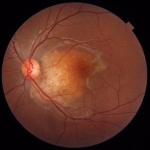
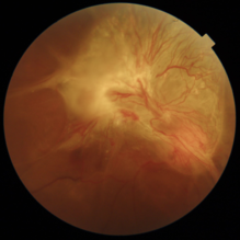
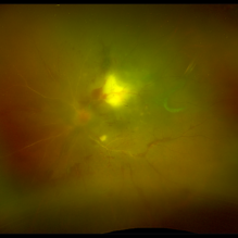
 The Dread of the Crimson Red
Jun 2 2025 by Thirumalesh Mochi Basavaraj, MD
Fundus photograph of a 64 year man post laser depicting a regressed NVD in the superior aspect and a Persistent Neo vascularization in the inferior aspect
Photographer: Vivek
Condition/keywords: Neovascularisation at the Disc (NVD), proliferative diabetic retinopathy (PDR)
-
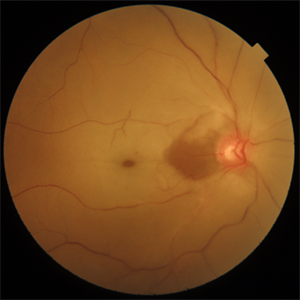
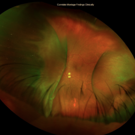
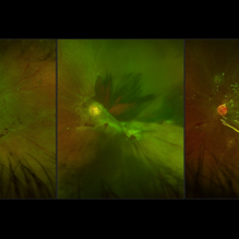
 The Headlight in the Fog
Jun 17 2025 by Thirumalesh Mochi Basavaraj, MD
37 year old male with sudden onset diminution of visual acuity has a large retinochoridal granuloma along the superotemporal arcade and a few with satellite lesions more temporal to it, there was extensive Occlusoive vasculitis (both arterioles and veins )being involved with Vitrities.
Photographer: Vivekanand ,Narayana nethralaya
Imaging device: Daytona
Condition/keywords: acute toxoplasmosis, retinochoroiditis
A project from the American Society of Retina Specialists