-
 Choroidal Nevus Color Fundus
Nov 13 2014 by Kathy Karsten, COT
Choroidal Nevus with cataract fragments in eye
Photographer: Kathy Karsten
Imaging device: Topcon TRC 50DX
Condition/keywords: choroidal nevus
-
 Melanocytic Choroidal Lesion Fundus Photo
Nov 13 2014 by Kathy Karsten, COT
Melanocytic choroidal lesion.
Photographer: Kathy Karsten
Imaging device: Topcon TRC 50DX
-
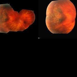
 Fundus Photos Malignant Melanoma
Nov 13 2014 by Kathy Karsten, COT
Malignant melanoma of eye.
Photographer: Kathy Karsten
Imaging device: Topcon TRC 50DX
Condition/keywords: malignant neoplasm of eye
-
 Fundus Photo Malignant Melanoma
Nov 13 2014 by Kathy Karsten, COT
Malignant melanoma.
Photographer: Kathy Karsten
Imaging device: Topcon TRC 50DX
Condition/keywords: melanoma
-
 Metastatic Malignant Melanoma
Nov 13 2014 by Kathy Karsten, COT
Metastatic malignant melanoma in 44-year-old man.
Photographer: Kathy Karsten
Imaging device: Topcon TRC 50DX
Condition/keywords: melanoma
-
 Retinoblastoma OD FA 6-2-2015-12-15-42 PM Proof
Jun 4 2015 by Kathy Karsten, COT
15-year-old male born with retinoblastoma. S/P enucleation OS at 3 months of age. S/P chemo/radiation lesions OD
Photographer: Kathy Karsten, COT
Imaging device: Topcon TRC 50-DX
Condition/keywords: retinoblastoma
-
 Malignant Melanoma
Jul 8 2015 by Kathy Karsten, COT
Fundus photograph of 70-year-old woman presenting with obscured vision in the right eye. Diagnosed with malignant melanoma. Patient underwent subsequent brachytherapy.
Photographer: Kathy Karsten, COT
Imaging device: Topcon TRC-50DX
-
 Ischemic Branch Retinal Vein Occlusion With Compensatory Collateral Vessels
Jul 8 2015 by Kathy Karsten, COT
Heidelberg fluorescein angiography picture of ischemic branch retinal vein occlusion with compensatory collateral vessels in 30-year-old woman.
Photographer: Kathy Karsten, COT
Imaging device: Heidelberg capturing system
Condition/keywords: ischemia
-
 Neovascular Glaucoma
Dec 4 2015 by Kathy Karsten, COT
Ahmad tube shunt with peaked pupil in the left eye of a 63-year-old woman for neovascular glaucoma.
Photographer: Kathy Karsten, COT
Imaging device: Topcon TRC 50-DX
Condition/keywords: angle neovascularization
-
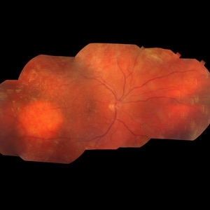
 Malignant Choroidal Melanoma
Dec 4 2015 by Kathy Karsten, COT
Malignant choroidal melanoma and branch retinal vein occlusion in 69-year-old male.
Photographer: Kathy Karsten, COT
Imaging device: Topcon TRC-50 DX
-
 Amelanotic Choroidal Lesion
Dec 4 2015 by Kathy Karsten, COT
Amelanotic choroidal lesion OD in 74-year-old male.
Photographer: Kathy Karsten, COT
Imaging device: Topcon TRC 50-DX
-
 Iris Nevus
Apr 11 2016 by Kathy Karsten, COT
Iris nevus in a 35-year-old woman.
Photographer: KATHY KARSTEN
Imaging device: TOPCON TRC-50DX
Condition/keywords: iris, nevus
-
 Vitreomacular Traction
Apr 11 2016 by Kathy Karsten, COT
OCT of vitreomacular traction in a 40-year-old man.
Photographer: KATHY KARSTEN
Imaging device: TOPCON TRC 50-DX
Condition/keywords: optical coherence tomography (OCT), vitreomacular membrane
-
 Iris Nevus
Apr 11 2016 by Kathy Karsten, COT
Anterior segment photo of an iris nevus with a peaked pupil observed over time for growth.
Photographer: KATHY KARSTEN
Imaging device: TOPCON 50-DX
Condition/keywords: iris, nevus
-
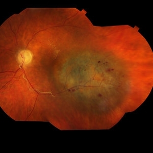
 Malignant Melanoma
Apr 11 2016 by Kathy Karsten, COT
Fundus photo montage of a malignant melanoma in a 70-year-old woman.
Photographer: KATHY KARSTEN, COT
Imaging device: TOPCON TRC 50 DX
Condition/keywords: malignant melanoma
A project from the American Society of Retina Specialists