-
 OCT Choroidal Tuberculoma
Jun 15 2020 by Aayesha Khanum
OCT choroidal tuberculoma.
Photographer: Puttaswamy
Imaging device: DRI OCT Triton swept source OCT-Topcon
Condition/keywords: abscess, choroidal tuberculoma
-
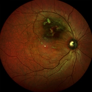
 Choroidal Tuberculoma
Jun 15 2020 by Aayesha Khanum
Fundus photo of diagnosed case of pulmonary TB.
Photographer: Puttaswamy
Imaging device: DRI OCT Triton swept source OCT- Topcon
Condition/keywords: abscess, choroidal tuberculoma
-
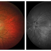 Regressed CRVO
Jun 27 2020 by Aayesha Khanum
Regressed CRVO with severe collateral at disc
Photographer: Ravikrishna, Puttaswamy
Imaging device: Heidelberg Spectralis
Condition/keywords: central retinal vein occlusion (CRVO), collaterals
-
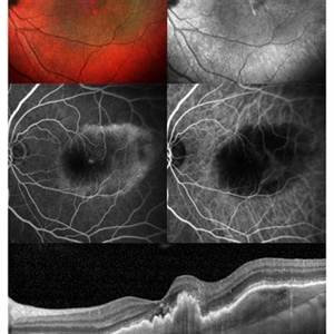 Neovascular AMD
Jun 27 2020 by Aayesha Khanum
Multicolor image and infra-red image showing fibrovascular pigment epithelial detachment (PED),FFA and ICG confirms the diagnosis., SD-OCT shows notched fibrovascular PED
Photographer: Puttaswamy,Ravikrishna
Imaging device: Heidelberg Spectralis
Condition/keywords: neovascular age-related macular degeneration (AMD)
-
 Supero-Temporal Branch Retinal Vein Occlusion
Jun 27 2020 by Aayesha Khanum
Fundus photograph showing branch retinal vein occlusion with macular edema.
Photographer: Ravikrishna, Puttaswamy
Imaging device: Heidelberg Spectralis
Condition/keywords: branch retinal vein occlusion (BRVO)
A project from the American Society of Retina Specialists