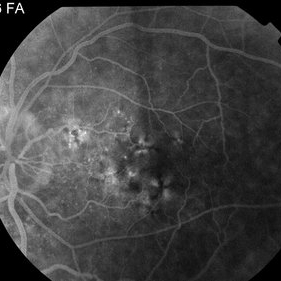
This 43 year woman has a history of myotonic dystrophy. Note the pigmentary changes in the macula typical of this disorder. Her visual acuity measured 20/30. The OCT was normal. She presented with a choroidal neovascular membrane in the fellow eye.
-
 Myotonic Dystrophy Maculopathy
Sep 12 2014 by Thomas A. Ciulla, MD, MBA, FASRS
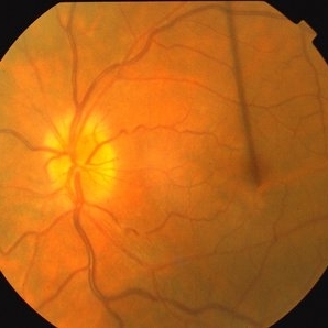
This 43-year-old woman has a history of myotonic dystrophy. Note the subtle pigmentary changes in the macula typical of this disorder. Her visual acuity measured 20/30.
Photographer: Thomas Steele
Condition/keywords: maculopathy
-
 Myotonic Dystrophy Maculopathy
Sep 12 2014 by Thomas A. Ciulla, MD, MBA, FASRS
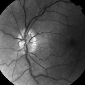
This 43-year-old woman has a history of myotonic dystrophy. Note the pigmentary changes in the macula typical of this disorder.
Photographer: Thomas Steele
Condition/keywords: maculopathy
-
 Myotonic Dystrophy Maculopathy
Sep 12 2014 by Thomas A. Ciulla, MD, MBA, FASRS
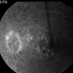
This 43-year-old woman has a history of myotonic dystrophy. Note the pigmentary changes in the macula typical of this disorder. There is no evidence of choroidal neovascularization in this left eye.
Photographer: Thomas Steele
Condition/keywords: maculopathy