A 48 year old woman with a history of optic nerve pit-related serous detachment of the macula previously underwent laser treatment at the temporal aspect of the optic nerve on the right. The serous detachment of the macula persisted. Since it had not responded, she was then told that she might have central serous retinopathy. She was referred to the retina service where she was noted to have visual acuity of 20/200 and persistent submacular fluid, felt to be due to the optic nerve pit. There was no evidence of a central serous retinopathy on angiography. OCT revealed submacular fluid with chronic features, including numerous large cystoid spaces. She underwent vitrectomy, additional laser treatment in the peripapillary region, and infusion of C3F8 gas with face-down positioning. The OCT show the sequential improvement of the macula at 1month, 3 months, 9 months, and 1 year. The visual acuity improved from 20/200 to 20/40 over that time period.
-
 Optic Nerve Pit Related Serous Retinal Detachment
Sep 15 2014 by Thomas A. Ciulla, MD, MBA, FASRS
Pre-op color fundus photograph showing optic nerve pit and prior laser treatment at temporal aspect of optic nerve.
Photographer: Thomas Steele
Condition/keywords: congenital optic nerve pit, pre-op, serous retinal detachment, vitrectomy, vitreomacular surgery
-
 Optic Nerve Pit Related Serous Retinal Detachment
Sep 15 2014 by Thomas A. Ciulla, MD, MBA, FASRS
Pre-op red-free photo showing optic nerve pit and prior laser treatment at temporal aspect of optic nerve. Note the subtle outline of the serous retinal detachment.
Photographer: Thomas Steele
Condition/keywords: congenital optic nerve pit, pre-op, red-free, serous retinal detachment, vitrectomy, vitreomacular surgery
-
 Optic Nerve Pit Related Serous Retinal Detachment
Sep 15 2014 by Thomas A. Ciulla, MD, MBA, FASRS
Pre-op early phase angiogram showing optic nerve pit and prior laser treatment at temporal aspect of optic nerve
Photographer: Thomas Steele
Condition/keywords: congenital optic nerve pit, pre-op, serous retinal detachment, vitrectomy, vitreomacular surgery
-
 Optic Nerve Pit Related Serous Retinal Detachment
Sep 15 2014 by Thomas A. Ciulla, MD, MBA, FASRS
Pre-op late phase angiogram showing optic nerve pit and prior laser treatment at temporal aspect of optic nerve
Photographer: Thomas Steele
Condition/keywords: congenital optic nerve pit, pre-op, serous retinal detachment, vitrectomy, vitreomacular surgery
-
 Optic Nerve Pit Related Serous Retinal Detachment
Sep 10 2014 by Thomas A. Ciulla, MD, MBA, FASRS
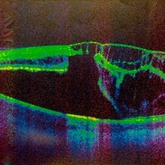
Pre-op OCT. The visual acuity measured 20/200. Note the severe subretinal fluid with chronic features, including the cystoid spaces.
Condition/keywords: congenital optic nerve pit
-
 Optic Nerve Pit Related Serous Retinal Detachment
Sep 8 2014 by Thomas A. Ciulla, MD, MBA, FASRS
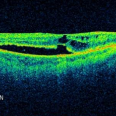
Post-op OCT at 1 month. Visual acuity improved to 20/80.
Condition/keywords: congenital optic nerve pit
-
 Optic Nerve Pit Related Serous Retinal Detachment
Sep 8 2014 by Thomas A. Ciulla, MD, MBA, FASRS
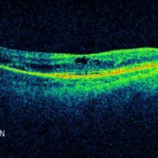
Post-op OCT at 3 months. Visual acuity measured 20/80.
Condition/keywords: congenital optic nerve pit
-
 Optic Nerve Pit Related Serous Retinal Detachment
Sep 8 2014 by Thomas A. Ciulla, MD, MBA, FASRS
Post-op OCT at 9 months. Visual acuity improved to 20/40.
Condition/keywords: congenital optic nerve pit
-
 Optic Nerve Pit Related Serous Retinal Detachment
Sep 8 2014 by Thomas A. Ciulla, MD, MBA, FASRS
Post-op OCT at 1 year. Visual acuity measured 20/40.
Condition/keywords: congenital optic nerve pit, post-op, serous retinal detachment, vitrectomy, vitreomacular surgery