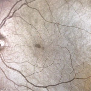
This patient presented with a distinct paracentral scotoma OS and a lesion that was difficult to visualize on color or red free photos. However, note the prominent wedge shaped lesion nasal to the fovea on the infrared photo, typical of acute macular neuroretinopathy. This case highlights the usefulness of the infrared photo for this condition.
-
 Acute Macular Neuroretinopathy
Sep 15 2014 by Thomas A. Ciulla, MD, MBA, FASRS
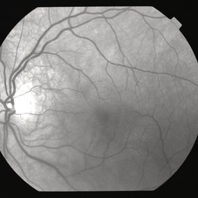
Color photo. This might be a typical fundus photo, with no definite lesion. However, the infrared photo nicely depicts a typical lesion.
Condition/keywords: acute macular neuroretinopathy, color photo
-
 Acute Macular Neuroretinopathy
Sep 15 2014 by Thomas A. Ciulla, MD, MBA, FASRS
Red-free photo. Similar to the color photo, this red-free photo shows no definite lesion. However, the infrared photo nicely depicts the typical lesion.
Condition/keywords: acute macular neuroretinopathy, red-free
-
 Acute Macular Neuroretinopathy
Aug 13 2014 by Thomas A. Ciulla, MD, MBA, FASRS
Infrared photo. This infrared photo nicely demonstrates a typical small wedge-shaped lesion of acute macular neuroretinopathy. This case highlights the usefulness of the infrared photo for this condition.
Condition/keywords: acute macular neuroretinopathy