-
 Myopic Foveoschisis
May 19 2014 by Ahmad Gawady
OCT Of 56-year-old highly myopic female with drop of vision BCVA 2/60. Clear media, myopic fundus , Ill defined macular abnormality. OCT shows inferior juxtafoveal splitting of Inner retinal layer. No evidence of PVD, CNV or leakage .
Condition/keywords: myopic foveoschisis, optical coherence tomography (OCT)
-
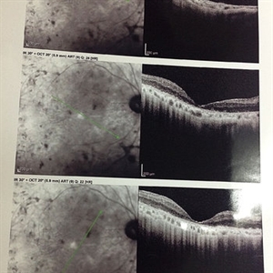 Myopic Foveoschisis
May 19 2014 by Ahmad Gawady
OCT Lt of a 56-year-old female with drop of vision OS bilateral high myopia -6.0 D. Normal I.O.P., BCVA 6/18 OD , 2/60 OS., clear media, myopic fundus OD. No abnormalities OD, myopic fundus Ill defined fovea abnormality OS. OCT macular area OS : inferior juxtafoveal splitting of inner retinal layer (myopic foveoschisis) No evidence of PVD, CNV or leakage.
Condition/keywords: high myopia, optical coherence tomography (OCT)
A project from the American Society of Retina Specialists