-
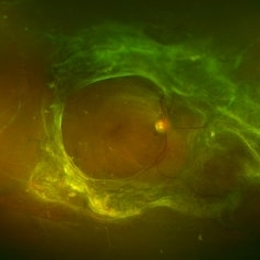
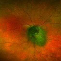 Nevus Turned Melanoma
May 9 2014 by Matt Poe, COA
The patient had a juxtapapillary nevus and did not return for two years until complaining of his right eye vision decreasing.
Photographer: Matt Poe COA, Northwest Arkansas Retina Associates, Springdale, AR.
Condition/keywords: choroidal nevus
-
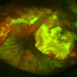
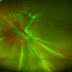
 Tractional Retinal Detachment
May 9 2014 by Matt Poe, COA
Proliferative diabetic retinopathy with tractional retinal detachment.
Photographer: Matt Poe COA, Northwest Arkansas Retina Associates, Springdale, AR.
Condition/keywords: proliferative diabetic retinopathy (PDR), tractional retinal detachment
-
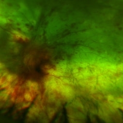
 Proliferative Diabetic Retinopathy
May 9 2014 by Matt Poe, COA
Proliferative diabetic retinopathy with capillary nonperfusion temporal to macula.
Photographer: Matt Poe COA, Northwest Arkansas Retina Associates, Springdale, AR.
Condition/keywords: proliferative diabetic retinopathy (PDR)
-
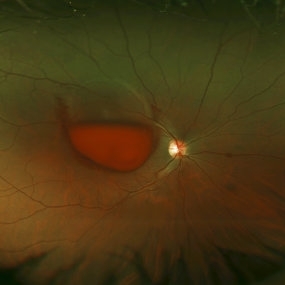
 Retinal Detachment
Dec 10 2014 by Matt Poe, COA
Old retinal detachment, patient only had LP out of that eye.
Photographer: Matt Poe, COA. Northwest Arkansas Retina Associates, Springdale, AR.
-
 Valsalva Retinopathy
Dec 12 2014 by Matt Poe, COA
A 21-year-old female presented with sudden decrease in VA. Patient explained that she had been sick and coughing a lot. Patient's VA was CF in that eye.
Photographer: Matt Poe, COA. Northwest Arkansas Retina Associates, Springdale, AR.
Condition/keywords: valsalva retinopathy
-
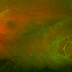 Paw Print Retinal Detachment
Dec 29 2014 by Matt Poe, COA
This patient had an inferior temporal retinal detachment that resembles a paw print.
Photographer: Matt Poe, COA. Northwest Arkansas Retina Associates, Springdale, AR.
-
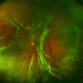 Funnel Retinal Detachment
Feb 2 2015 by Matt Poe, COA
The patient presented with total vision loss for >2months. Patient had history of exudative ARMD with intravitreal injections. No surgical intervention was done due to the long standing detachment and patient health.
Photographer: Matt Poe, COA. Northwest Arkansas Retina Associates, Springdale, AR.
Condition/keywords: retinal defect
-
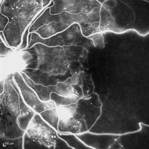
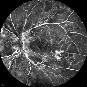
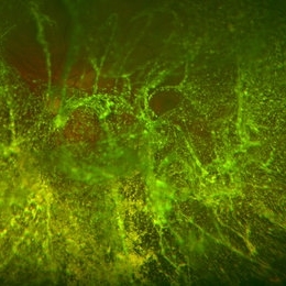
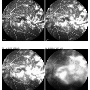 Proliferative Diabetic Retinopathy with Neovascular Membrane
Feb 2 2015 by Matt Poe, COA
IVFA of a 34-year-old lady diagnosed with proliferative diabetic retinopathy with neovascular membrane.
Photographer: Matt Poe, COA. Northwest Arkansas Retina Associates, Springdale, AR.
Imaging device: Heidelberg HRA
Condition/keywords: neovascular membrane, proliferative diabetic retinopathy (PDR)
-
 Pigmentary Retinal Dystrophy
Feb 9 2015 by Matt Poe, COA
This was a lady that presented with bilateral pigmentary retinal dystrophy.
Photographer: Matt Poe, COA. Northwest Arkansas Retina Associates, Springdale, AR.
Condition/keywords: hereditary retinal dystrophy, pigmentary retinal dystrophy
-
 Tractional Retinal Detachment
Feb 9 2015 by Matt Poe, COA
This patient presented with bilateral tractional retinal detachments secondary to her proliferative diabetic retinopathy. Surprisingly the patient had 20/60 in that eye.
Photographer: Matt Poe, COA. Northwest Arkansas Retina Associates, Springdale, AR.
Condition/keywords: diabetic mellitus, proliferative diabetic retinopathy (PDR), tractional retinal detachment
-
 TRD Secondary to PDR
Feb 10 2015 by Matt Poe, COA
This patient has a long standing tractional detachment secondary to his proliferative diabetic retinopathy. Patient is CF in this eye.
Photographer: Matt Poe, COA. Northwest Arkansas Retina Associates, Springdale, AR.
Imaging device: Heidelberg HRA
Condition/keywords: diabetes, proliferative diabetic retinopathy (PDR), red-free, tractional retinal detachment
-
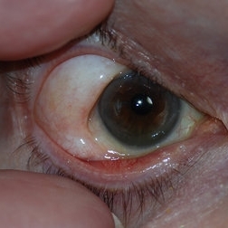
 Scleral Buckle Protrusion
Feb 23 2015 by Matt Poe, COA
This was taken with a Nikon D80 camera to document the protrusion of the scleral buckle.
Photographer: Matt Poe, COA. Northwest Arkansas Retina Associates, Springdale, AR.
Condition/keywords: external, extruded scleral buckle, scleral buckle
-
 Herniated Orbital Fat
Feb 23 2015 by Matt Poe, COA
This was taken with a Nikon D80 camera to document the herniated orbital fat.
Photographer: Matt Poe, COA. Northwest Arkansas Retina Associates, Springdale, AR.
Condition/keywords: herniated orbital fat, orbital fat invasion
-
 Retinal Fold
Mar 9 2015 by Matt Poe, COA
This patient developed a retinal fold following a retinal detachment repair. The patient underwent another retinal detachment surgery to fix the retinal fold. The patient's retina was fixed and did well post-operative.
Photographer: Matt Poe, COA. Northwest Arkansas Retina Associates, Springdale, AR.
Condition/keywords: optical coherence tomography (OCT), retinal fold
-
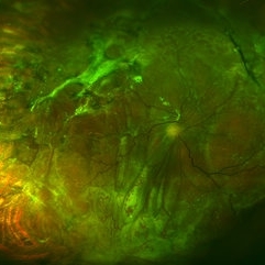
 Proliferative Diabetic Retinopathy
Mar 16 2015 by Matt Poe, COA
IVFA of 53-year-old male with Proliferative Diabetic Retinopathy, Diabetic macular edema, and a tractional retinal detachment.
Photographer: Matt Poe, COA. Northwest Arkansas Retina Associates, Springdale, AR.
Imaging device: Heidelberg HRA
Condition/keywords: neovascularization (NV), proliferative diabetic retinopathy (PDR)
-
 Proliferative Diabetic Retinopathy Inverted
May 28 2015 by Matt Poe, COA
This is a young man with advanced proliferative diabetic retinopathy.
Photographer: Matt Poe, COA. Northwest Arkansas Retina Associates, Springdale, AR.
Imaging device: Heidelberg HRA
Condition/keywords: capillary dropouts, diabetes, neovascularization elsewhere (NVE), proliferative diabetic retinopathy (PDR)
-
 Old Retinal Detachment
Aug 3 2015 by Matt Poe, COA
This is a fundus photo of a young man in his 20s with a long-standing retinal detachment.
Photographer: Matt Poe, COA. Northwest Arkansas Retina Associates, Springdale, AR.
Condition/keywords: retinal fibrosis
-
 Total Retinal Detachment
Sep 21 2015 by Matt Poe, COA
This patient has a long standing total retinal detachment. His vision at the time of the visit was NLP.
Photographer: Matt Poe, COA. Northwest Arkansas Retina Associates, Springdale, AR.
Condition/keywords: blind eye
-
 Panuveits / Disseminated Chorioretinitis
Nov 24 2015 by Matt Poe, COA
This is of a 42-year-old woman that presented to our office with visual acuity of NLP. Labs show that patient is positive for cytomegalovirus. Labs with increased IgG titer for CMV, but normal IgM. She has been diagnosed with Panuveitis, Disseminated chorioretinitis, and serous retinal detachment.
Photographer: Matt Poe, COA. Northwest Arkansas Retina Associates, Springdale, AR.
Condition/keywords: panuveitis, serous retinal detachment
-
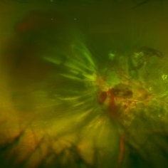
 Extreme Asteroid Hyalosis
Apr 27 2016 by Matt Poe, COA
This patient was sent for a possible retinal detachment. Extreme difficult view of posterior pole due to asteroid hyalosis. After B-Scan was performed it was determined patient did not have a retinal detachment, only posterior vitreous detachment.
Photographer: Matt Poe, COA. Northwest Arkansas Retina Associates, Springdale, AR.
Condition/keywords: asteroid hyalosis, posterior vitreous detachment
A project from the American Society of Retina Specialists