-
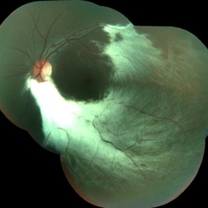
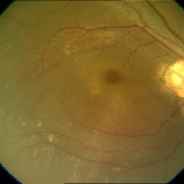 Disc Pit With Maculopathy
Jun 3 2014 by Neha Goel, MS DNB FRCS (Glasg)
Fundus photograph of the right eye of a 28-year-old male.
Photographer: Neha Goel
Imaging device: Zeiss Visucam
Condition/keywords: congenital optic nerve pit, neurosensory detachment of retina
-
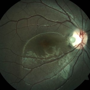
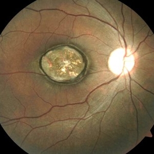
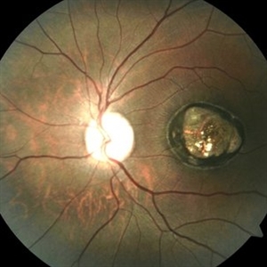
 Disc Drusen
Jun 3 2014 by Neha Goel, MS DNB FRCS (Glasg)
Fundus photograph of the left eye of a 48-year-old male.
Photographer: Neha Goel
Imaging device: Zeiss Visucam
Condition/keywords: drusen of optic disc
-
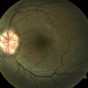
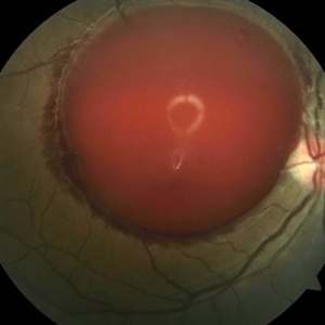
 Optic Disc Pit
Jun 3 2014 by Neha Goel, MS DNB FRCS (Glasg)
Fundus photograph of the right eye of a 15-year-old male.
Photographer: Neha Goel
Imaging device: Zeiss Visucam
Condition/keywords: optic disc pit
-
 Epiretinal Membrane
Aug 31 2014 by Neha Goel, MS DNB FRCS (Glasg)
Fundus photograph of a 32-year-old female.
Photographer: Neha Goel
Imaging device: Zeiss Visucam
Condition/keywords: epiretinal membrane (ERM), epiretinal membrane formation, idiopathic epiretinal membrane
-
 Choroidal Rupture
Apr 27 2015 by Neha Goel, MS DNB FRCS (Glasg)
Fundus photograph of the left eye of a 32-year-old male following blunt trauma with a ball.
Photographer: Neha Goel
Imaging device: Zeiss visucam
Condition/keywords: blunt trauma, choroidal rupture, subretinal hemorrhage
-
 Subhyaloid Hemorrhage
Apr 29 2015 by Neha Goel, MS DNB FRCS (Glasg)
Fundus photograph of the right eye of a 25-year-old male with complaints of loss of vision following a bout of coughing.
Photographer: Neha Goel
Imaging device: Zeiss visucam
Condition/keywords: subhyaloid hemorrhage, valsalva retinopathy
-
 Subhyaloid Hemorrhage - Post Hyaloidotomy
Apr 29 2015 by Neha Goel, MS DNB FRCS (Glasg)
Fundus photograph immediately following Nd:YAG laser hyaloidotomy.
Photographer: Neha Goel
Imaging device: Zeiss visucam
Condition/keywords: hyaloidotomy, subhyaloid hemorrhage, valsalva retinopathy
-
 Myelinated nerve fibres
Apr 29 2015 by Neha Goel, MS DNB FRCS (Glasg)
Montage fundus photograph of the left eye of a 16-year-old boy.
Photographer: Neha Goel
Imaging device: Zeiss visucam
Condition/keywords: myelinated nerve fiber layer, myelinated nerve fibers
-
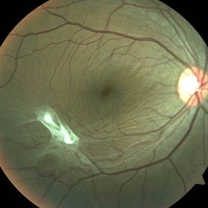
 Congenital Toxoplasmosis RE
Apr 29 2015 by Neha Goel, MS DNB FRCS (Glasg)
Fundus photograph of the right eye of a 25-year-old male with decreased vision since early childhood.
Photographer: Neha Goel
Imaging device: Zeiss visucam
Condition/keywords: congenital toxoplasmosis, inactive toxoplasmosis, toxoplasmosis
-
 Congenital Toxoplasmosis LE
Apr 29 2015 by Neha Goel, MS DNB FRCS (Glasg)
Fundus photograph of the left eye of a 25-year-old male with decreased vision since early childhood.
Photographer: Neha Goel
Imaging device: Zeiss visucam
Condition/keywords: congenital toxoplasmosis, inactive toxoplasmosis, toxoplasmosis
A project from the American Society of Retina Specialists