-
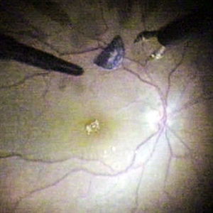 Intraocular Foreign Body (IOFB)
Apr 17 2014 by Shlomit Schaal, MD, PhD, MHCM
Intra-operative photograph of a metal foreign body being removed with intraocular foreceps.
Photographer: Shlomit Schaal MD, PhD and Nathan Podoll MD, University of Louisville, Louisville, KY
Condition/keywords: intraocular foreign body
-
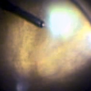 Endogenous Candida Endophthalmitis
Apr 17 2014 by Shlomit Schaal, MD, PhD, MHCM
Intraoperative photo of vitrectomy for endogenous endopthalmitis.
Photographer: Shlomit Schaal MD, PhD and Nathan Podoll MD, University of Louisville, Louisville, KY
Condition/keywords: endogenous endophthalmitis
-
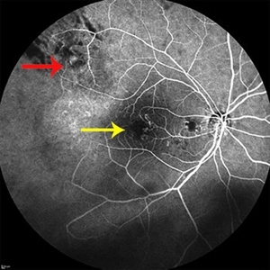 Radiation Retinopathy
Jun 5 2014 by Shlomit Schaal, MD, PhD, MHCM
Angiogram showing previously irradiated and regressed peripheral choroidal melanoma (red arrow), and the resulting central radiation retinopathy with retinal ischemia and remodeling of capillaries (yellow arrow).
Photographer: Shlomit Schaal MD, PhD, University of Louisville, Louisville, KY
Condition/keywords: radiation retinopathy
-
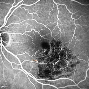 RAM
RAM
Jun 5 2014 by Shlomit Schaal, MD, PhD, MHCM
Fundus angiogram showing 171 microns retinal arterial macroaneurysm that previously bled and now remains throbmbotic.
Photographer: Shlomit Schaal MD, PhD, University of Louisville, Louisville, KY
Condition/keywords: retinal arterial macroaneurysm
-
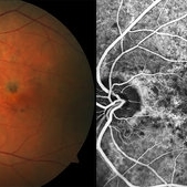 Angioid Streaks
Jun 5 2014 by Shlomit Schaal, MD, PhD, MHCM
Color photo (left) and angiogram (right) of a patient with choroidal angioid streaks who subsequently developed a subfoveal choroidal neovascular membrane.
Photographer: Shlomit Schaal MD, PhD, University of Louisville, Louisville, KY
Condition/keywords: angioid streaks
-
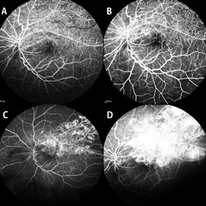 VVA
VVA
Jun 5 2014 by Shlomit Schaal, MD, PhD, MHCM
Fundus angiograms showing chronic ischemic branch retinal retinal vein occlusion with venous-venous anastomosis formation.
Photographer: Shlomit Schaal MD, PhD and Shivani Reddy MD, University of Louisville, Louisville, KY
Condition/keywords: venous-venous anastomosis
-
 Foreign Body SS
Feb 14 2015 by Shlomit Schaal, MD, PhD, MHCM
A four millimeter metal foreign body removed surgically using foreign body forceps. The macula is protected by PFCL heavy fluid. There is a traumatic retinal detachment inferiorly (top of photo, surgeon's view).
Photographer: Shlomit Schaal, MD, PhD
Condition/keywords: intraocular foreign body
A project from the American Society of Retina Specialists