-
 CNV Due to Toxoplasmosis
Apr 6 2014 by Ratimir Lazic, MD, PhD
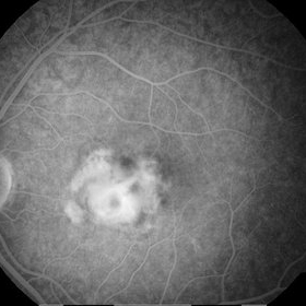
A FAG image of a 7-year-old boy. Late venous phase image shows unsharply limited leakage of dye which presents staining of chorioretinal scar with active CNV.
Photographer: Marko Vlasic, University Eye Clinic Svjetlost
Imaging device: Zeis Visucam Lite 2
Condition/keywords: choroidal neovascularization (CNV), toxoplasmosis
-
 CNV due to Toxoplasmosis
Apr 6 2014 by Ratimir Lazic, MD, PhD
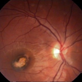
A color fundus image of a 7-year-old boy. Pigmented chorioretinal scar sorrounded by subretinal hemorrhage can be seen. VA is 0,2 by Snellen lines. The image presents the baseline clinical picture. The antiVEGF intravitreal injection, under general anesthesia, was administered.
Photographer: Marko Vlasic, University Eye Clinic Svjetlost
Imaging device: Zeis Visucam Lite 2
Condition/keywords: choroidal neovascularization (CNV), subretinal hemorrhage, toxoplasmosis
-
 CNV due to Toxoplasmosis
Apr 6 2014 by Ratimir Lazic, MD, PhD
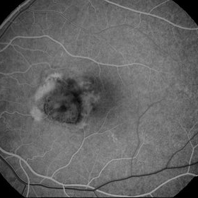
A FAG image of a 7-year-old boy. On early venous phase image can be seen hypoflorescent area in foveal region with a leakege of dye.
Photographer: Marko Vlasic, University Eye Clinic Svjetlost
Imaging device: Zeis Visucam Lite 2
Condition/keywords: choroidal neovascularization (CNV), toxoplasmosis
A project from the American Society of Retina Specialists