-
 Branch Retinal Vein Occlusion
Jul 21 2020 by Patrik Rajs
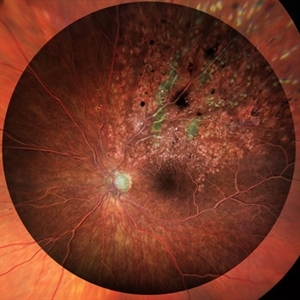
Combination of Color and FAG image of a patient with a branch retinal vein occlusion after laser treatment.
Photographer: Patrik Rajs, EYE CLINIC of Jan Evangelista Purkyne University and Masaryk Hospital in Usti nad Labem, Czech Republic
Condition/keywords: branch retinal vein occlusion (BRVO)
-
 Rhegmatogenous Retinal Detachment
Mar 3 2021 by Patrik Rajs
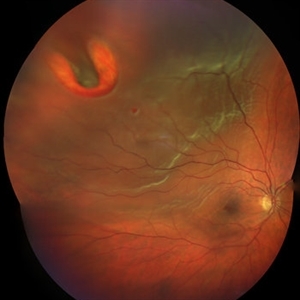
A 51-year-old female patient presented with inferior nasal scotoma and 5/10 vision in the right eye due to a retinal detachment with a giant retinal horseshoe tear.
Photographer: Patrik Rajs, EYE CLINIC of Jan Evangelista Purkyne University and Masaryk Hospital, Czech Republic, Ústí nad Labem
Imaging device: Clarus 700
Condition/keywords: giant retinal tear
-
 Choroid Detachment
Jul 7 2021 by Patrik Rajs
This eye was a tough one. The patient underwent PPV twice, the second one with silicone oil (SO) for retinal re-detachment. Due to the development of secondary glaucoma, silicone oil evacuation and lavage of the anterior chamber were performed. Because of the high IOP even after the evacuation, the XEN was implanted. The surgery was followed by choroidal detachment presented in the picture on the left side along with the residual silicone bubble superiorly. The retinal tear is captured inferiorly surrounded by laser spots. The second image (on the right) was taken only 7 days later and it shows that choroidal detachment in the eye resolved completely.
Photographer: Patrik Rajs, EYE CLINIC of Jan Evangelista Purkyne University and Masaryk Hospital, Czech Republic, Ústí nad Labem
Condition/keywords: choroid, detachment, glaucoma, retina, silicone oil, tear
A project from the American Society of Retina Specialists