-
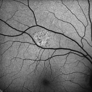
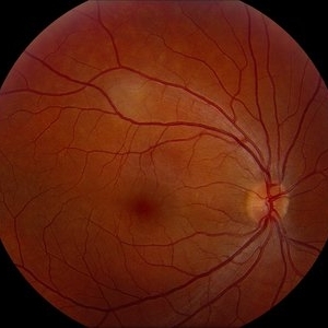 Unilateral Acute Idiopathic Maculopathy - Color Photo
Aug 26 2012 by Robin Ray, MD
Fundus photograph of a 32-year-old woman with several day history of inferior scotoma in the right eye. Vision in both eyes is 20/20. No vitreous cell in either eye. Has a history of hand, foot, and mouth disease 7-10 days prior to presentation. A diagnosis of unilateral acute idiopathic maculopathy was made.
Photographer: Kidron Robertson, Georgia Eye Institute of the Southeast, Savannah, GA
Condition/keywords: chorioretinal inflammations, Coxsackie, unilateral acute idiopathic maculopathy
-
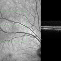
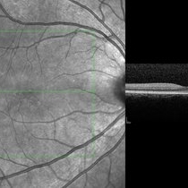 Unilateral Acute Idiopathic Maculopathy - Infrared Photo with OCT through fovea
Aug 26 2012 by Robin Ray, MD
Infrared photo shows subtle RPE changes on inferior edge of fovea. OCT does not show any significant RPE or IS/OS disruption.
Photographer: Kidron Robertson, Georgia Eye Institute of the Southeast, Savannah, GA
Imaging device: Heidelberg Spectralis
Condition/keywords: chorioretinal inflammations, Coxsackie, unilateral acute idiopathic maculopathy
-
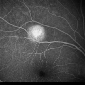 Unilateral Acute Idiopathic Maculopathy - Fluorescein Angiography (2)
Sep 9 2012 by Robin Ray, MD
FA of UAIM (late) - hyperintense staining of lesion.
Photographer: Kidron Robertson, Georgia Eye Institute of the Southeast, Savannah, GA
Imaging device: Heidelberg Spectralis
Condition/keywords: chorioretinal inflammations, Coxsackie, unilateral acute idiopathic maculopathy
-
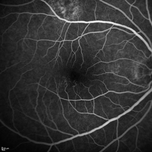 Unilateral Acute Idiopathic Maculopathy - Fluorescein Angiography (1)
Sep 9 2012 by Robin Ray, MD
Early recirculation phase showing stippled hyperfluorescence of inferior fovea without leakage. Lesion superiorly shows heterogeneous fluorescence.
Photographer: Kidron Robertson, Georgia Eye Institute of the Southeast, Savannah, GA
Imaging device: Heidelberg Spectralis
Condition/keywords: chorioretinal inflammations, Coxsackie, unilateral acute idiopathic maculopathy
-
 Unilateral Acute Idiopathic Maculopathy
Sep 9 2012 by Robin Ray, MD
Image on right shows infrared photo of the lesion of interest. SDOCT (image on left) through the lesion of interest shows hyperreflectivity of the RPE with disruption/loss of detail of the IS/OS junction.
Photographer: Kidron Robertson - Georgia Eye Institute of the Southeast
Imaging device: Heidelberg Spectralis
Condition/keywords: chorioretinal inflammations, Coxsackie, unilateral acute idiopathic maculopathy
-
 Unilateral Acute Idiopathic Maculopathy - Fundus Autofluorescence
Sep 9 2012 by Robin Ray, MD
FAF of UAIM lesion
Photographer: Kidron Robertson, Georgia Eye Institute of the Southeast, Savannah, GA
Imaging device: Heidelberg Spectralis
Condition/keywords: chorioretinal inflammations, Coxsackie, unilateral acute idiopathic maculopathy
A project from the American Society of Retina Specialists