-
 Capillary Hemangioma
Dec 14 2016 by Young Hee Yoon, MD, PhD
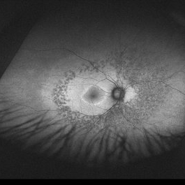
Wide fundus photo of a 35-year-old man with huge capillary hemagioma in the right eye. He is diagnosed with Von Hippel-Lindau disease. His best-corrected visual acuity was 20/50.
Photographer: Yu Jin Jang and Hun Eui Hong, Asan Medical Center
Imaging device: Wide fundus camera
Condition/keywords: Von Hippel-Lindau
-
 Capillary Hemangioma
Dec 14 2016 by Young Hee Yoon, MD, PhD
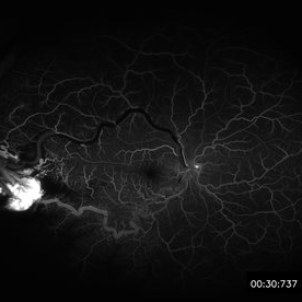
Wide fluorescein angiography of a 35-year-old man with huge capillary hemagioma in the right eye. He is diagnosed with Von Hippel-Lindau disease. His best-corrected visual acuity was 20/50.
Photographer: Yu Jin Jang and Hun Eui Hong, Asan Medical Center
Imaging device: Wide fluorescein angiography
Condition/keywords: Von Hippel-Lindau
-
 Capillary Hemangioma
Dec 14 2016 by Young Hee Yoon, MD, PhD
Wide fluorescein angiography of a 35-year-old man with huge capillary hemagioma in the right eye. He is diagnosed with Von Hippel-Lindau disease. His best-corrected visual acuity was 20/50.
Photographer: Yu Jin Jang and Hun Eui Hong, Asan Medical Center
Imaging device: Wide fluorescein angiography
Condition/keywords: Von Hippel-Lindau
-
 Choroidal Neovascularization in Patient With Choroidal Osteoma
Dec 14 2016 by Young Hee Yoon, MD, PhD
Fundus photography of a 56-year-old woman with choroidal neovascularization at the margin of the choroidal osteoma in the right eye. Her best-corrected visual acuity was 20/30.
Photographer: Soon Tae Kim, Asan Medical Center
Condition/keywords: choroidal neovascularization (CNV), choroidal osteoma
-
 Choroidal Neovascularization in Patient With Choroidal Osteoma
Dec 14 2016 by Young Hee Yoon, MD, PhD
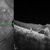
Spectral domain optical coherence tomography of a 56-year-old woman with choroidal neovascularization at the margin of the choroidal osteoma in the right eye. Her best-corrected visual acuity was 20/30.
Photographer: Mi Hwa Choi, Asan Medical Center
Condition/keywords: choroidal neovascularization (CNV), choroidal osteoma
-
 Choroidal Neovascularization in Patient With Choroidal Osteoma
Dec 14 2016 by Young Hee Yoon, MD, PhD
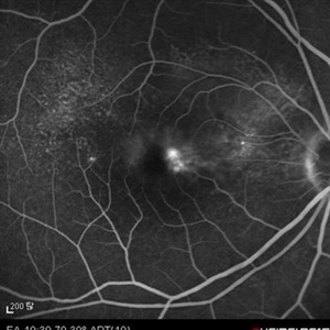
Fluorescein angiography of a 56-year-old woman with choroidal neovascularization at the margin of the choroidal osteoma in the right eye. Her best-corrected visual acuity was 20/30.
Photographer: Soon Tae Kim, Asan Medical Center
Condition/keywords: choroidal neovascularization (CNV), choroidal osteoma
-
 Choroidal Neovascularization in Patient With Choroidal Osteoma
Dec 14 2016 by Young Hee Yoon, MD, PhD
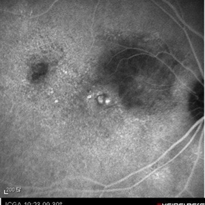
Indocyanine green angiography of a 56-year-old woman with choroidal neovascularization at the margin of the choroidal osteoma in the right eye. Her best-corrected visual acuity was 20/30.
Photographer: Soon Tae Kim, Asan Medical Center
Condition/keywords: choroidal neovascularization (CNV), choroidal osteoma
-
 Welder's Maculopathy
Dec 14 2016 by Young Hee Yoon, MD, PhD
Early phase fluorescein angiography of a 53-year-old man with welder's maculopathy in the right eye. He complained sudden visual loss in the right eye at thirty minutes after welding arc work. His best-corrected visual acuity was hand motion.
Photographer: Kyung Wun Kim, Asan Medical Center
Condition/keywords: central retinal artery occlusion (CRAO), Welder's maculopathy
-
 Welder's Maculopathy
Dec 14 2016 by Young Hee Yoon, MD, PhD
Late phase fluorescein angiography of a 53-year-old man with welder's maculopathy in the right eye. He complained sudden visual loss in the right eye at thirty minutes after welding arc work. His best-corrected visual acuity was hand motion.
Photographer: Kyung Wun Kim, Asan Medical Center
Condition/keywords: central retinal artery occlusion (CRAO), Welder's maculopathy
-
 Welder's Maculopathy
Dec 14 2016 by Young Hee Yoon, MD, PhD
Fundus photograph of a 53-year-old man with welder's maculopathy in the right eye. He complained sudden visual loss in the right eye at thirty minutes after welding arc work. His best-corrected visual acuity was hand motion.
Photographer: Kyung Wun Kim, Asan Medical Center
Condition/keywords: central retinal artery occlusion (CRAO), Welder's maculopathy
-
 Pericentral HCQ (hydroxychloroquine) Toxicity
Dec 21 2016 by Young Hee Yoon, MD, PhD
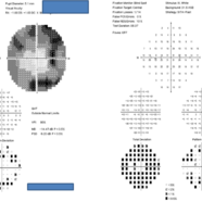
Visual field of an 47-year-old woman with pericentral HCQ (hydroxychloroquine) toxicity (both eyes).
Photographer: Da Jung Kim, Asan Medical Center
Imaging device: Visual field
Condition/keywords: pericentral HCQ (hydroxychloroquine) toxicity
-
 Pericentral HCQ (hydroxychloroquine) Toxicity
Dec 21 2016 by Young Hee Yoon, MD, PhD
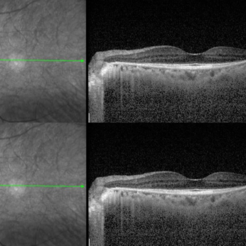
OCT (optical coherence tomography) of an 47-year-old woman with pericentral HCQ (hydroxychloroquine) toxicity (Left eye).
Photographer: Jung Im Jo, Asan Medical Center
Condition/keywords: optical coherence tomography (OCT), pericentral HCQ (hydroxychloroquine) toxicity
-
 Pericentral HCQ (hydroxychloroquine) Toxicity
Dec 21 2016 by Young Hee Yoon, MD, PhD
OCT (optical coherence tomography) of an 47-year-old woman with pericentral HCQ (hydroxychloroquine) toxicity (Right eye).
Photographer: Jung Im Jo, Asan Medical Center
Condition/keywords: optical coherence tomography (OCT), pericentral HCQ (hydroxychloroquine) toxicity
-
 Pericentral HCQ (hydroxychloroquine) Toxicity
Dec 21 2016 by Young Hee Yoon, MD, PhD
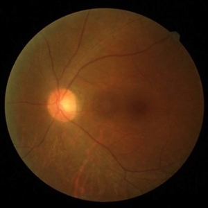
Fundus photograph of an 47-year-old woman with pericentral HCQ (hydroxychloroquine) toxicity (left eye).
Photographer: Hyae Jin Jo, Asan Medical Center
Condition/keywords: pericentral HCQ (hydroxychloroquine) toxicity
-
 Pericentral HCQ (hydroxychloroquine) Toxicity
Dec 21 2016 by Young Hee Yoon, MD, PhD
Fundus photograph of an 47-year-old woman with pericentral HCQ (hydroxychloroquine) toxicity (right eye).
Photographer: Hyae Jin Jo, Asan Medical Center
Condition/keywords: pericentral HCQ (hydroxychloroquine) toxicity
-
 Pericentral HCQ (hydroxychloroquine) Toxicity
Dec 21 2016 by Young Hee Yoon, MD, PhD
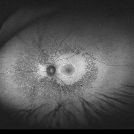
Wide auto-fluorescence photograph of an 47-year-old woman with pericentral HCQ (hydroxychloroquine) toxicity (left eye).
Photographer: Kyung Woon Kim, Asan Medical Center
Imaging device: Wide Auto fluorescence photograph
Condition/keywords: pericentral HCQ (hydroxychloroquine) toxicity
-
 Pericentral HCQ (hydroxychloroquine) Toxicity
Dec 21 2016 by Young Hee Yoon, MD, PhD
Wide auto-fluorescence photograph of an 47-year-old woman with pericentral HCQ (hydroxychloroquine) toxicity (right eye).
Photographer: Kyung Woon Kim, Asan Medical Center
Imaging device: Wide Auto fluorescence photograph
Condition/keywords: pericentral HCQ (hydroxychloroquine) toxicity
-
---thumb.jpg/image-square;max$300,300.ImageHandler) Ocular Lymphoma
Ocular Lymphoma
Jan 3 2014 by Young Hee Yoon, MD, PhD
Ultrasonographic image of a 51-year-old female in a complete remission status after treatment for diffuse large B cell lymphoma (DLBCL) (Stage IV). Her best-corrected visual acuity was 20/80. Cytology for vitreous fluid revealed malignant lymphocytes, suggesting vitreoretinal relapse.
Photographer: Kyoung Woon Kim, Asan Medical Center
Condition/keywords: ocular lymphoma
-
---thumb.jpg/image-square;max$300,300.ImageHandler) Ocular Lymphoma
Ocular Lymphoma
Jan 3 2014 by Young Hee Yoon, MD, PhD
Wide field fundus photograph of a 51-year-old female in a complete remission status after treatment for diffuse large B cell lymphoma (DLBCL) (Stage IV). Her best-corrected visual acuity was 20/80. Cytology for vitreous fluid revealed malignant lymphocytes, suggesting vitreoretinal relapse.
Photographer: Soo Hyun Cho, Asan Medical Center
Imaging device: Fundus photography using Optomap, optos
Condition/keywords: ocular lymphoma
A project from the American Society of Retina Specialists