-
 Tilted Disc Syndrome Complicated with RPE Atrophy and Polypoidal Choroidal Vasculopathy
Jan 20 2020 by Pierre-Henry Gabrielle, MD
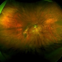
Fundus optos photograph of an 81-year-old woman with a tilted disc syndrome complicated with RPE atrophy and polypoidal choroidal vasculopathy.
Photographer: Pierre-Henry Gabrielle, Ophthalmology department, Dijon University Hospital, France.
Imaging device: Optos
Condition/keywords: atrophic pigment epithelium, indocyanine green (ICG) angiography, polypoidal choroidal vasculopathy (PCV), tilted disc
-
 Tilted Disc Syndrome Complicated with RPE Atrophy and Polypoidal Choroidal Vasculopathy
Jan 20 2020 by Pierre-Henry Gabrielle, MD
Zoomed fundus optos photograph of an 81-year-old woman with a tilted disc syndrome complicated with RPE atrophy and polypoidal choroidal vasculopathy.
Photographer: Pierre-Henry Gabrielle, Ophthalmology department, Dijon University Hospital, France.
Imaging device: Optos
Condition/keywords: atrophic pigment epithelium, Optos, polypoidal choroidal vasculopathy (PCV), tilted disc
-
 Tilted Disc Syndrome Complicated with RPE Atrophy and Polypoidal Choroidal Vasculopathy
Jan 20 2020 by Pierre-Henry Gabrielle, MD
Coupled OCT B-scan and ICG angiography of an 81-year-old woman with a tilted disc syndrome complicated with RPE atrophy and polypoidal choroidal vasculopathy.
Photographer: Pierre-Henry Gabrielle, Ophthalmology department, Dijon University Hospital, France.
Imaging device: Heidelberg spectralis
Condition/keywords: atrophic pigment epithelium, indocyanine green (ICG) angiography, optical coherence tomography (OCT), polypoidal choroidal vasculopathy (PCV), tilted disc
-
 Tilted Disc Syndrome Complicated with RPE Atrophy and Polypoidal Choroidal Vasculopathy
Jan 20 2020 by Pierre-Henry Gabrielle, MD
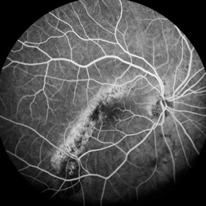
Early-phase FA angiography of an 81-year-old woman with a tilted disc syndrome complicated with RPE atrophy and polypoidal choroidal vasculopathy.
Photographer: Pierre-Henry Gabrielle, Ophthalmology department, Dijon University Hospital, France.
Imaging device: Heidelberg spectralis angiography
Condition/keywords: atrophic pigment epithelium, polypoidal choroidal vasculopathy (PCV), tilted disc
-
 Tilted Disc Syndrome Complicated with RPE Atrophy and Polypoidal Choroidal Vasculopathy
Jan 20 2020 by Pierre-Henry Gabrielle, MD
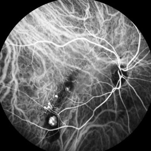
Early-phase ICG angiography of an 81-year-old woman with a tilted disc syndrome complicated with RPE atrophy and polypoidal choroidal vasculopathy.
Photographer: Pierre-Henry Gabrielle, Ophthalmology department, Dijon University Hospital, France.
Imaging device: Heidelberg spectralis angiography
Condition/keywords: atrophic pigment epithelium, indocyanine green (ICG) angiography, polypoidal choroidal vasculopathy (PCV), tilted disc
-
 Tilted Disc Syndrome Complicated with RPE Atrophy and Polypoidal Choroidal Vasculopathy
Jan 20 2020 by Pierre-Henry Gabrielle, MD
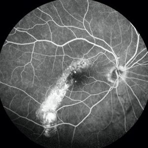
Late-phase fluorescein angiography of an 81-year-old woman with a tilted disc syndrome complicated with RPE atrophy and polypoidal choroidal vasculopathy.
Photographer: Pierre-Henry Gabrielle, Ophthalmology department, Dijon University Hospital, France.
Imaging device: Heidelberg spectralis angiography
Condition/keywords: atrophic pigment epithelium, polypoidal choroidal vasculopathy (PCV), tilted disc
-
 Tilted Disc Syndrome Complicated with RPE Atrophy and Polypoidal Choroidal Vasculopathy
Jan 20 2020 by Pierre-Henry Gabrielle, MD
Late-phase ICG angiography of an 81-year-old woman with a tilted disc syndrome complicated with RPE atrophy and polypoidal choroidal vasculopathy.
Photographer: Pierre-Henry Gabrielle, Ophthalmology department, Dijon University Hospital, France.
Imaging device: Heidelberg spectralis angiography
Condition/keywords: atrophic pigment epithelium, indocyanine green (ICG) angiography, polypoidal choroidal vasculopathy (PCV), tilted disc
A project from the American Society of Retina Specialists