Search results (88 results)
-
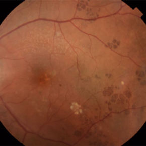
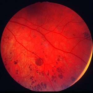
 Benign Lobular Inner Nuclear Layer Proliferations (BLIP)
Apr 15 2024 by Virginia Gebhart
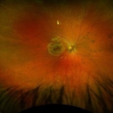
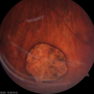
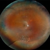
29 year old male with multiple flat CHRPE lesions, genetic testing negative for ACP genes associated with Gardner syndrome. Multiple intraretinal amelanotic lesions consistent with Benign Lobular Inner Nuclear Layer Proliferations (BLIP) of the retina
Photographer: Virginia Gebhart
Imaging device: Topcon
Condition/keywords: BLIP Benign Lobular Inner Nuclear Layer Proliferations, CHRPE, congenital hypertrophy of the retinal pigment epithelium (CHRPE)
-
 CHRPE
CHRPE
Jan 6 2025 by Kavitha Duraipandi, MD DNB FICO FRCS
Bear tracks (animal tracks, grouped pigmentation spots) are simply many small CHRPEs located in isolated small area of the retina. These have not been reported to have the potential to transform into adenocarcinoma but yearly evaluations may be prudent.
Condition/keywords: CHRPE
-
 CHRPE
CHRPE
Mar 25 2025 by Toolie Winters
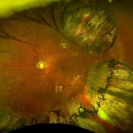
Ultra-wide field fundus photograph of a 78-year-old woman with extensive CHRPE lesions OS. Continued observation has been recommended at this time.
Photographer: Toolie Winters
Imaging device: Optos California
Condition/keywords: CHRPE, congenital hypertrophy of the retinal pigment epithelium (CHRPE), fundus photography, Optos, Optos California, pseudocolor, ultra-wide field imaging
-
 CHRPE
CHRPE
Apr 1 2019 by Gary R. Cook, MD, FACS
35 year-old white female with asymptomatic CHRPE lesion OS; V.A.= 20/20-2.
Imaging device: Topcon VT-50
Condition/keywords: congenital hypertrophy of the retinal pigment epithelium (CHRPE)
-
 CHRPE
CHRPE
May 9 2014 by S. Natarajan, MD, FASRS, FRCS (GLASGOW) , FICO, D.Sc, FELA
23-year-old male in for routine eye checkup with BCVA 6/6 (OU). Showed CHRPE lesion in INQ (OD )on fundus.
Photographer: ADITYA JYOT EYE HOSPITAL,MUMBAI,INDIA
Condition/keywords: congenital hypertrophy of the retinal pigment epithelium (CHRPE), myopic eye
-
 CHRPE
CHRPE
Dec 13 2018 by Jason Griffith
50-year-old male being followed for typical CHRPE presentation.
Photographer: Jason Griffith, Tennessee Retina, Nashville, TN
Imaging device: Clarus 500
Condition/keywords: congenital hypertrophy of the retinal pigment epithelium (CHRPE)
-
 CHRPE
CHRPE
Jun 3 2020 by stephen oconnell
Congenital hypertrophy of the RPE with minimal pigment
Condition/keywords: congenital hypertrophy of the retinal pigment epithelium (CHRPE)
-
 CHRPE
CHRPE
Jun 5 2020 by stephen oconnell
CHRPE
Condition/keywords: congenital hypertrophy of the retinal pigment epithelium (CHRPE)
-
 CHRPE
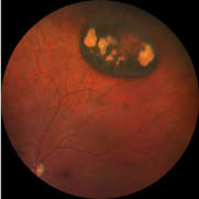
CHRPE
Jan 12 2023 by Christopher R. Adam, M.D.
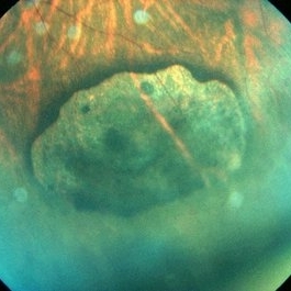
Optos color photograph of a 51 y/o F with a 9x9mm CHRPE in the nasal quadrant. The lesion is flat with a bordering halo and lacunae.
Condition/keywords: congenital hypertrophy of the retinal pigment epithelium (CHRPE)
-
 CHRPE
CHRPE
-
 CHRPE
CHRPE
Oct 8 2019 by DIEGO TOLENTINO
CHRPE plus laser barricade around retinal break
Photographer: Diego Tolentino
Condition/keywords: congenital hypertrophy of the retinal pigment epithelium (CHRPE)
-
 CHRPE
CHRPE
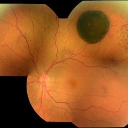
Jan 15 2021 by Priya Rasipuram Chandrasekaran, MBBS, DO, DNB, FRCS
This is the fundus photo and fundus photo montage of the left eye of a 25-year-old male showing flat, solitary, round, greyish pigmented lesion situated AT THE equator with a scalloped margin. Vessels overlying the lesion are normal and there is a clear demarcation line between this and normal retina. The margins are hypopigmented with few hypopigmented lacunae inside.
Condition/keywords: congenital hypertrophy of the retinal pigment epithelium (CHRPE)
-
 CHRPE - "BEAR TRACKS" PATTERN
Nov 8 2022 by Heitor Nogueira
BEAR TRACKS
Photographer: Heitor Nogueira
Condition/keywords: bear tracks, CHRPE, congenital hypertrophy of the retinal pigment epithelium (CHRPE)
-
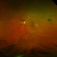
 CHRPE and Bear Tracks
Jan 7 2025 by Drew Mitchell
Fundus Autofluorescence of a CHRPE and Bear Tracks.
Photographer: Drew Mitchel, OCT-C
Imaging device: Optos Silverstone
Condition/keywords: bear tracks, CHRPE, congenital hypertrophy of the retinal pigment epithelium (CHRPE)
-
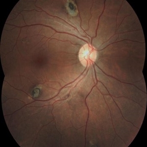
 Gardner's Syndrome
Nov 10 2023 by Virginia Gebhart
17-year-old male with multiple pigmented spots most likely related to Gardner's Syndrome. Pt has not been diagnosed with FAP at this time, however pt receives regular screenings. Extensive maternal family hx of FAP syndrome and colon cancer. Pt's mother has FAP, who had colon resection. Pt's 2 aunts, 1 uncle, grandmother and great grandmother all passed from colon cancer. Pt has multiple maternal cousins diagnosed with FAP
Photographer: Virginia Gebhart
Imaging device: Topcon
Condition/keywords: CHRPE, congenital hypertrophy of the retinal pigment epithelium (CHRPE), familial adenomatous polyposis, Gardner Syndrome
-
 Gardner's Syndrome (FAP)
Nov 3 2023 by Virginia Gebhart
43 year-old female with multiple CHRPE lesions consistent with Gardner's Syndrome. Patient had colectomy at age 21. Patient is one of 7 children, 6 had FAP, 3 had colon removal and are alive and well, 3 have passed away from colon cancer. Maternal mother and grandmother also passed away from colon cancer. Patient has 2 children, one son (17) also has Gardner's Syndrome (FAP)
Photographer: Virginia Gebhart
Imaging device: Topcon
Condition/keywords: CHRPE, Gardner Syndrome
-
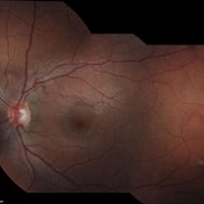
 Lattice Degeneration With Atrophic Retinal Holes
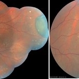
Jan 30 2025 by Kimberly Wakester
Ultra-wide field montage fundus photograph of a 56-year-old woman with lattice degeneration with atrophic holes statues post laser. Patient also has a small CHRPE temporal to macula and trace ERM that is not visually significant. Will continue follow up care to monitor and treat as needed.
Photographer: Kimberly Wakester, COA
Imaging device: Optos California
Condition/keywords: atrophic retinal hole, CHRPE, epiretinal membrane (ERM), lattice degeneration, montage photo
-
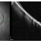 OCT Over Congenital Hypertrophy of the Retinal Pigment Epithelium (CHRPE)
Mar 25 2022 by Elaine Michele Binkley, MD
Optical coherence tomography image over a CHRPE lesion.
Photographer: Meghan Menzel, University of Iowa
Condition/keywords: CHRPE
-
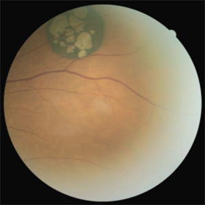 Solitary Congenital Hypertrophy of the Retinal Pigment Epithelium with Lacunae
Jun 11 2023 by Ethan K Sobol, MD
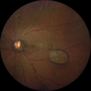
Fundus photograph of a solitary CHRPE in the superotemporal quadrant with central hypopigmented lacunae.
Condition/keywords: CHRPE, congenital hypertrophy of the retinal pigment epithelium (CHRPE), lacunae
-
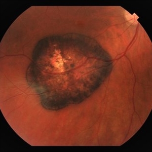
 Solitary large Congenital Hypertrophy of Retinal Pigment Epithelium (CHRPE)
Jul 1 2023 by Aditya S Kelkar, MS, FRCS, FASRS,FRCOphth
Right eye fundus photograph of a 42 year old asymptomatic male demonstrating a superotemporal solitary large Congenital Hypertrophy of Retinal Pigment Epithelium (CHRPE) lesion.
Photographer: Optom Komal Jangam
Imaging device: OPTOS DAYTONA
Condition/keywords: CHRPE
-
 CHRPE & Myelinated RNFL
May 21 2020 by John S. King, MD
47-year-old white female, asymptomatic, sent to evaluate a scar OD. 20/40 cc, normotensive, examination significant for a flat, solitary lesion with pigmented borders and depigmented center with early lacunae forming, along with myeliated RNFL at the temporal edge of the lesion.
Photographer: Kay Dalby
Imaging device: Topcon
Condition/keywords: congenital hypertrophy of the retinal pigment epithelium (CHRPE), myelinated nerve fiber layer
-
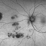
 CHRPE - grouped pigmentation
Jan 11 2013 by Alex P. Hunyor, MD
Congenital grouped pigmentation of the RPE ("bear tracks").
Condition/keywords: bear tracks, congenital hypertrophy of the retinal pigment epithelium (CHRPE)
-
 CHRPE and CHRRPE
May 26 2024 by shama sharief
Funds photo with CHRPE and CHRRPE.
Condition/keywords: CHRRPE, congenital hypertrophy of the retinal pigment epithelium (CHRPE)
-
 Congenital Hypertrophy of the Retinal Pigment Epithelium (CHRPE)
Aug 24 2012 by Andrew N. Antoszyk, MD FASRS
CHRPE lesion (black pigmented lesion) located along superior temporal arcade of left eye
Photographer: Lorainne Clark, Charlotte Eye Ear Nose and Throat Associates
-
 61-Year-Old Man With Large Peripheral CHRPE
Dec 9 2017 by Timothy S Fuller, MD
61-year-old man presented for evaluation of pigmented retinal lesion. Found to have a large, peripheral CHRPE with characteristic lacunae, sharp margins, and lack of elevation.
Condition/keywords: benign pigmented lesions, congenital hypertrophy of the retinal pigment epithelium (CHRPE), lacunae
 Loading…