Search results (477 results)
-
 Retinal capillary hemangiomas 3
Jan 11 2013 by Alex P. Hunyor, MD
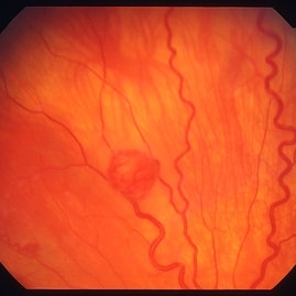
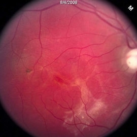
Retinal capillary haemangiomas, left superior periphery, in a 20 year old female with von Hippel-Lindau disease.
Condition/keywords: hemangioma, retinal capillary hemangioma, Von Hippel-Lindau
-
 Retinal Angiomatous Proliferation in Age-Related Macular Degeneration with Subretinal Neovascularization
Sep 24 2012 by James B. Soque, CRA, OCT-C, COA, FOPS
75-year-old white male with classic SRN with RAP. Lesion OD is active, and patient is receiving anti-VEGF treatment. Mid phase FA, 50 Deg, Mag 2x.
Photographer: James Soque, CRA, COA, Island Retina, Shirley, NY, USA
Imaging device: Topcon TRC 50 DX, OIS 5.0 MP Color, FA Camera, OIS Software
Condition/keywords: age-related macular degeneration (AMD), fundus autofluorescence (FAF), leakage, retinal angiomatous proliferation (RAP), subretinal neovascularization (SRNV)
-
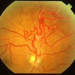
 Racemose Hemangioma
Feb 20 2013 by From the Collections of Thomas M. Aaberg, MD and Thomas M. Aaberg Jr., MD
Dilated tortuous blood vessels.
Condition/keywords: racemose hemangioma
-
 Von Hippel-Lindau
Sep 3 2012 by Hamid Ahmadieh, MD
Color fundus photograph of a 35-year-old woman with retinal angiomatosis.
Photographer: Hamid Ahmadieh, MD, Ophthalmic Research Center, Labbafinejad Medical Center, Shahid Beheshti University of Medical Sciences , Tehran
Imaging device: Topcon Fundus Camera
Condition/keywords: retinal angiomatous proliferation (RAP), Von Hippel-Lindau
-
 Retinal capillary hemangioma 2
Jan 11 2013 by Alex P. Hunyor, MD
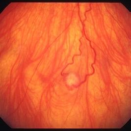
Retinal capillary haemangioma, right inferior periphery, in a 20-year-old female with von Hippel-Lindau disease.
Condition/keywords: hemangioma, retinal capillary hemangioma, Von Hippel-Lindau
-
 Sturge-Weber Episcleral-Vessels
Apr 17 2014 by Susanna S. Park, MD, PhD
External photo of the right eye of this 8-year-old Hispanic boy with Sturge -Weber Syndrome and diffuse choroidal hemangioma showing dilated episcleral vessels.
Photographer: Ellen Redenbo, University of California Davis Eye Center
Condition/keywords: dilated episcleral vessels, Sturge-Weber syndrome
-
 Von Hippel-Lindau
Sep 3 2012 by Hamid Ahmadieh, MD
Color fundus photograph of a 35-year-old woman with retinal angiomatosis.
Photographer: Hamid Ahmadieh, MD, Ophthalmic Research Center, Labbafinejad Medical Center, Shahid Beheshti University of Medical Sciences
Imaging device: Topcon Fundus Camera
Condition/keywords: retinal angiomatous proliferation (RAP), Von Hippel-Lindau
-
 Retinal Angiomas In VHL
Dec 24 2012 by Roy D. Brod, MD
Fundus photograph of 16 year old male with recent diagnosis of Von Hippel-Lindau disease showing typical appearance of a retinal angioma in superior mid periphery OD. Note unrelated choroidal nevus above superior arcade.
Photographer: Julia Walker
Condition/keywords: hemangioma, Von Hippel-Lindau
-
 Von Hippel-Lindau
Sep 5 2012 by Hamid Ahmadieh, MD
Color fundus photograph of a 32-year-old man with retinal angiomatosis.
Photographer: Hamid Ahmadieh, MD, Ophthalmic Research Center, Labbafinejad Medical Center, Shahid Beheshti University of Medical Sciences
Imaging device: Topcon Fundus Camera
Condition/keywords: retinal angiomatous proliferation (RAP), Von Hippel-Lindau
-
---thumb.jpg/image-square;max$300,300.ImageHandler) Sturge-Weber Diffuse Hemangioma and Retinal Detachment on B-scan
Sturge-Weber Diffuse Hemangioma and Retinal Detachment on B-scan
Apr 18 2014 by Susanna S. Park, MD, PhD
B-scan ultrasonogram of the right eye of an 8 year old Hispanic boy with Sturge -Weber Syndrome showing diffuse choroidal thickening from diffuse choroidal hemangioma and associated total exudative retinal detachment.
Photographer: Ellen Redenbo, University of California Davis Eye Center
Condition/keywords: B scan ultrasound, diffuse choroidal hemangioma, Sturge-Weber syndrome
-
 Von Hippel-Lindau
Sep 8 2012 by Hamid Ahmadieh, MD
Color fundus photograph of a 32-year-old man with retinal angiomatosis.
Photographer: Hamid Ahmadieh, MD, Ophthalmic Research Center, Labbafinejad Medical Center, Shahid Beheshti University of Medical Sciences
Imaging device: Topcon Fundus Camera
Condition/keywords: retinal angiomatous proliferation (RAP), Von Hippel-Lindau
-
 Capillary Hemongima, Coat's Response
May 2 2013 by Henry J. Kaplan, MD
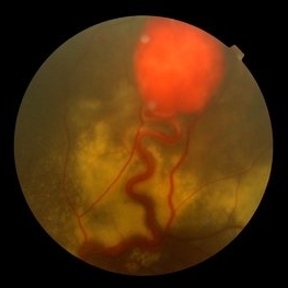
Coat's response as exudation in the macula in the same patient with retinal capillary hemangioma. Notice the dilated feeder vessles from the optic nerve infriorly; #2.
Condition/keywords: Coats' disease, retinal capillary hemangioma, Von Hippel-Lindau
-
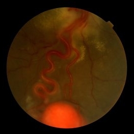
 Wyburn Mason Syndrome
May 2 2013 by Henry J. Kaplan, MD
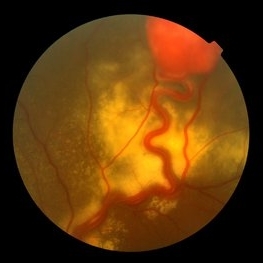
Racemose angioma of the retina in Wyburn Mason syndrome.
Condition/keywords: racemose hemangioma
-
 Diffuse Choroidal Hemangioma
Nov 7 2012 by Rajiv Anand, MD, FRCS, FASRS
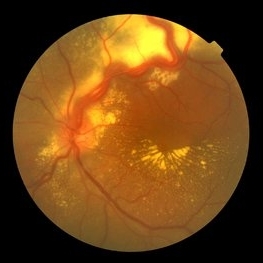
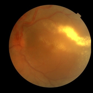
Fundus photo shows classic 'tomato-ketchup' red appearance of diffuse hemangioma. Due to chronic SRF , there is subretinal fibrosis.
Condition/keywords: subretinal fibrosis
-
 Von Hippel-Lindau
Oct 13 2012 by Hamid Ahmadieh, MD
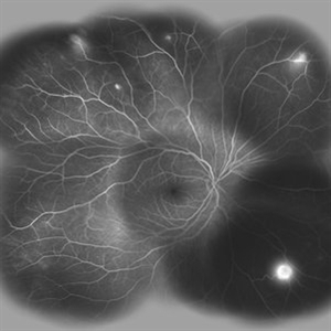
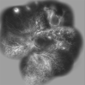
Wide field FA image of the right eye of a 25-year-old woman with retinal angiomatosis (Von Hippel-Lindau). Fundus of the right eye seemed to be normal in ophthalmoscopy.
Photographer: Soodabeh Fooladin, Negah Eye Center, Tehran
Imaging device: Heidelberg Spectralis
Condition/keywords: exudative retinal detachment, retinal angiomatous proliferation (RAP), Von Hippel-Lindau
-
 Von Hippel-Lindau 1
Oct 13 2012 by Hamid Ahmadieh, MD
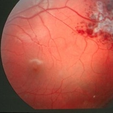
Color fundus photograph of the left eye of a 25-year-old woman with exudative retinal detachment secondary to retinal angiomatosis (Von Hippel-Lindau).
Photographer: Hamid Ahmadieh, MD, Ophthalmic Research Center, Labbafinejad Medical Center, Shahid Beheshti University of Medical Sciences
Imaging device: Topcon Fundus Camera
Condition/keywords: exudative retinal detachment, retinal angiomatous proliferation (RAP), Von Hippel-Lindau
-
 Retinal capillary hemangioma
Jan 11 2013 by Alex P. Hunyor, MD
Retinal capillary haemangioma nasal to optic disc, right eye.
Condition/keywords: retinal capillary hemangioma, Von Hippel-Lindau
-
 Von Hippel-Lindau
Sep 3 2012 by Hamid Ahmadieh, MD
Color fundus photograph of a 35-year-old woman with retinal angiomatosis.
Photographer: Hamid Ahmadieh, MD, Ophthalmic Research Center, Labbafinejad Medical Center, Shahid Beheshti University of Medical Sciences
Imaging device: Topcon Fundus Camera
Condition/keywords: retinal angiomatous proliferation (RAP)
-
 Choroidal Hemangioma
Oct 20 2012 by Hyung-Woo Kwak, MD
Fundus, ICG, and OCT examination showed a typical chorioretinal scar lying concentric to the optic disc. Typical choroidal rupture was performed after intravitreal gas injection under the diagnosis of submacular hemorrhage caused by trauma, after the absorption of submacular hemorrhage
Condition/keywords: chorioretinal scar, choroidal rupture, submacular hemorrhage
-
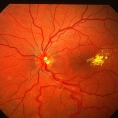
 Retinal Angiomas In VHL
Dec 24 2012 by Roy D. Brod, MD
Fundus photograph of 16 year old male with recent diagnosis of Von Hippel-Lindau disease showing 2 retinal angiomas in inferior mid periphery OD.
Photographer: Julia Walker
Condition/keywords: hemangioma, Von Hippel-Lindau
-
 Von Hippel-Lindau
Oct 13 2012 by Hamid Ahmadieh, MD
Wide field FA image of the left eye of a 25-year-old woman with exudative retinal detachment secondary to retinal angiomatosis (Von Hippel-Lindau).
Photographer: Soodabeh Fooladin, Negah Eye Center, Tehran
Imaging device: Heidelberg Spectralis
Condition/keywords: exudative retinal detachment, retinal angiomatous proliferation (RAP), Von Hippel-Lindau
-
---thumb.jpg/image-square;max$300,300.ImageHandler) Retinal capillary hemangioma 4 image 1
Retinal capillary hemangioma 4 image 1
Jan 11 2013 by Alex P. Hunyor, MD
Retinal capillary haemangioma, left eye, in a young female with von Hippel-Lindau disease. Color image 1 showing extensive lipid deposition in the macula.
Condition/keywords: retinal capillary hemangioma, Von Hippel-Lindau
-
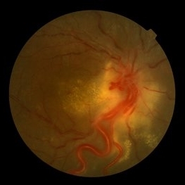
 Wyburn-Mason (Raecemose Hemangioma)
Oct 2 2013 by Jerald A. Bovino, MD
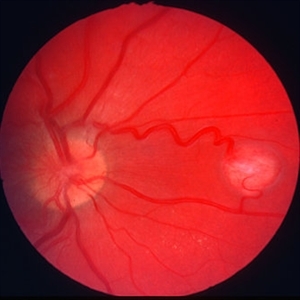
The patieint has typical Wyburn Mason syndrome with dilated, extrement tortuous venules.
Condition/keywords: dilated and tortous veins, racemose hemangioma
-
 Cavernous Hem of Retina
Oct 9 2012 by Alan D. Letson, MD
12-year-old boy with cavernous hemangioma of the retina.
Photographer: Beverly Radcliffe
Condition/keywords: cavernous hemangioma of the retina, hamartoma, phakoma
-
 Retinal Angiomas In VHL
Dec 24 2012 by Roy D. Brod, MD
Mid phase fluorescein angiogram of 16 year old male with recent diagnosis of Von Hippel-Lindau disease showing hyperfluorescent angioma in superior mid periphery OD.
Photographer: Julia Walker
Condition/keywords: hemangioma, Von Hippel-Lindau
 Loading…