Search results (905 results)
-
 Retinitis Pigmentosa
Apr 1 2025 by Jordyn Beckman
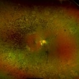
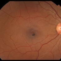
63 year old woman with Retinitis Pigmentosa observed over time with peripheral loss. Over the span of 5 years BCVA changed from 20/25 to 20/50.
Photographer: Jordyn Beckman, Retina Consultants of Carolina, P.A.
Imaging device: Optos California
Condition/keywords: atrophy, bone spicules, retinitis pigmentosa
-
 Shunt Vessels
Apr 1 2025 by Korey Starkey
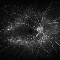
62-year-old patient presented with stable CRVO in the left eye. FA performed that day shows delayed AV transit is present, this has compensated with shunt vessels at the disc. However there is no evidence of active leakage. OS vision 20/25.
Photographer: Korey Starkey
Imaging device: Topcon
Condition/keywords: central retinal vein occlusion (CRVO), fundus photograph, optic nerve, shunts vessels, Topcon
-
 Ozurdex in AC
Apr 1 2025 by Korey Starkey
90-year-old patient with an Ozurdex implant that migrated into the AC and with the cornea decompensating. Patient recommended for urgent surgery to remove implant. Vision OD at this visit was CF @ 2ft, most recent visit vision is 20/400, PH 20/25.
Photographer: Korey Starkey
Imaging device: Topcon
Condition/keywords: anterior chamber, corneal decompensation, external, external photography, Ozurdex implant, Topcon
-
 Suspicious Nevus
Jan 15 2025 by Virginia Gebhart
14 year female with suspicious nevus located adjacent to the optic nerve. Questionable orange pigment present and worsening SRF compared to previous photos/OCT. RPE atrophy also present from previous fluid. No elevation. Will continue observation. BCVA 20/25
Photographer: Virginia Gebhart, Retina Consultants of Carolina
Imaging device: Topcon 50DX
Condition/keywords: choroidal nevus, nevus
-
 Pattern dystrophies – OCT demonstrates significant RPE irregularities and multiple focal inner segment-outer segment (IS-OS) disruptions with overlying cystic changes in both eyes of a 60-year-old man with PD
Sep 17 2024 by Nicolas A Yannuzzi, MD
Visual acuity was 20/20 OD and 20/25 OS. Genetic testing showed only 1 pathogenic variation in ABCA4, which is atypical for STGD1 Stargardt disease that is inherited in autosomal recessive fashion. (Images courtesy of Byron L. Lam, MD)
Condition/keywords: inherited retinal disease, pattern dystrophy
-
 Central Retinal Vein Occlusion with Macular Edema OS
Jul 5 2024 by Zach Seim
Optos fundus photograph of a Central Retinal Vein Occlusion in a 20 year old male. Vision at presentation was Dsc 20/25-1.
Photographer: Zach Seim
Imaging device: Optos California
Condition/keywords: central retinal vein occlusion (CRVO), left eye, macular edema, Optos, OPTOS CALIFORNIA
-
 Geographic Atrophy
Apr 22 2024 by Angela Rico
59 year-old female with MM1 Mitochondrial Genetic Defect. V/A- OD: 20/25, OS:20/40
Photographer: Angela Rico M.D.
Condition/keywords: Dystrophy of the Retinal Pigment Epithelium
-
 Advanced Stargardt Disease
Mar 25 2024 by Angela Rico
69 yr old female who presents with VA: OD 20/CF@4', OS 20/250.
Photographer: Angela Rico M.D., Retina Specialists of Tampa
Condition/keywords: Stargardts Disease
-
 Solar Retinopathy
Mar 17 2024 by Hector Gabriel Moreno Solano, MD, MHA
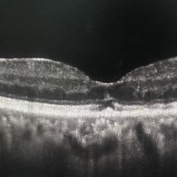
OCT scan of a 65 year old male with a history of direct exposure to solar eclipse rays, visual acuity of affected eye 20/80, contralateral eye 20/25.
Photographer: Héctor Gabriel Moreno-Solano, MD, MHA
Imaging device: Revo Optopol
Condition/keywords: light toxicity, macula, solar retinopathy
-
 Central Retinal Vein Occlusion with Macular Edema in Antiphospholipid Syndrome
Dec 24 2023 by Nikhil K Bommakanti, MD
A man in his thirties presented with a central retinal vein occlusion with macular edema in the right eye. Vision improved from 20/70 to 20/25 after 1 treatment with intravitreal bevacizumab. Laboratory testing revealed the presence of lupus anticoagulant.
Condition/keywords: antiphospholipid antibody syndrome, central retinal vein occlusion (CRVO), cystoid macular degeneration, macular edema
-
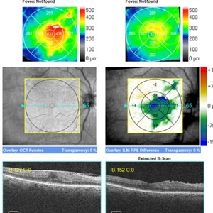
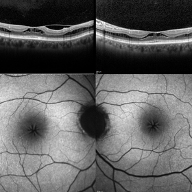 Macular Foveoschisis
Nov 9 2023 by Charlotte Jones
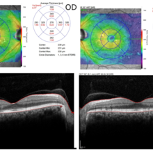
Bilateral ocular coherence tomography and fundus autofluorescence of a 77 year old woman with Macular Foveoschisis. Patient with stable vision since her last appointment (20/30 right eye and 20/25 left eye) with worsening vitreomacular traction in the right eye. Patient is followed routinely for Plaquenil use.
Photographer: Charlotte Jones
Imaging device: Heidelberg Spectralis
Condition/keywords: fundusautofluorescence, macularfoveoschisis, macularretinoschisis, macularstar, ocularcoherencetomography
-
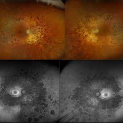 Retinitis Pigmentosa
Nov 7 2023 by Jolee Rodriguez
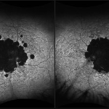
Bilateral fundus photography and fundus autofluorescence imaging of a 62-year-old male with Retinitis Pigmentosa. Patient reported visual field defects and dark adapting issues. Patient's vision at the time images were taken were sc20/20 of the right eye and sc20/25 of the left eye. Dr. Sutherland determined that based on the patient's lack of family history, the most likely route of inheritance is autosomal recessive.
Photographer: Jolee Rodriguez
Imaging device: Optos California RGB
Condition/keywords: autofluorescence imaging, fundus photography, hereditary retinal dystrophy, Optos, OPTOS CALIFORNIA RGB, retinitis pigmentosa, ultra-wide field imaging, Ultra-wide field retinal imaging, ultra-widefield image
-
 Congenital Retinal Macrovessel
Oct 13 2023 by Jacob D. Grodsky, MD
41 y/o male who presented with acute onset of blurred vision OD. Visual acuity was 20/200 OD; 20/25 OS. Examination was consistent with congenital retinal macrovessel through the macula with intraretinal hemorrhage as seen in the fundus photo. Intravitreal bevacizumab was injected, and visual acuity improved to 20/40 at 4-week follow-up. MRA head and neck was ordered to rule out other vascular anomalies.
Condition/keywords: congenital retinal macrovessel, RETINAL MACROVESSEL
-
 Neovascularization in Posterior Uveitis
Jul 27 2023 by Zach Seim
An ultra-widefield fluorescein angiogram of a 72 year old male with Posterior Uveitis and Neovascularization affecting the right eye. Patient's vision at the time of the image was Dcc 20/25. Dr. Korot states that the fluorescein angiogram shows patchy leakage throughout both eyes, with peripheral nonperfusion and secondary neovascularization. The patient was asked to get an extensive serological workup in an effort to identify any systemic autoimmune or infectious etiology as the cause for their severe inflammation.
Photographer: Zach Seim
Imaging device: OPTOS California
Condition/keywords: fluorescein angiogram (FA), FLUORESCEIN ANGIOGRAPHY, fluorescein leakage, neovascularization (NV), Optos, OPTOS CALIFORNIA, posterior uveitis, right eye, ultra-wide field imaging, ultra-widefield image
-
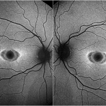 Bilateral “Bull's eye”pattern maculopathy
Mar 14 2023 by Anfisa Ayalon, MD
Both eyes fundus autofluorescence image of a 38-year-old female with “Bull's eye” pattern maculopathy. There is no history of medication use associated with retinal toxicity. BCVA RE 20/25+2, LE 20/20-3
Photographer: Danielle Ferguson and Alec Bertoni, University of Pittsburgh Medical Center
Condition/keywords: bull's eye maculopathy, maculopathy, retina
-
 "The Eye of Sauron"
Mar 14 2023 by Anfisa Ayalon, MD
Fundus autofluorescence image of a 38-year-old female with “Bull's eye” pattern maculopathy. There is no history of medication use associated with retinal toxicity. BCVA RE 20/25+2
Photographer: Danielle Ferguson and Alec Bertoni, University of Pittsburgh Medical Center
Condition/keywords: bull's eye maculopathy, retina
-
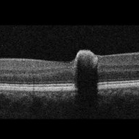 Congenital Simple Hamartoma of the Retinal Pigment Epithelium
May 16 2022 by David C Sousa, MD PhD FRANZCO
A 49-year-old man was referred after an incidental finding in the right eye macula. Best-corrected visual acuity was 20/25. Anterior segment examination was unremarkable. Fundoscopy revealed a juxta-foveal heavily pigmented well-demarcated slightly elevated lesion measuring 0.4 x 0.4 mm. No other changes were observed adjacent to the lesion or elsewhere in either eye. Optical coherence tomography revealed an area of retinal elevation with high optical reflectivity and posterior shadowing. The findings are consistent with congenital simple hamartoma of the retinal pigment epithelium. Given the benign, non-progressive and usually asymptomatic nature of this condition, most patients are diagnosed in adulthood.
Imaging device: Topcon Maestro2
Condition/keywords: retinal pigment epithelium (RPE) hamartoma
-
 Peripheral Drusen
Jan 19 2022 by Olivia Rainey
Ultra-widefield fluorescein angiogram of an 83-year-old female with Peripheral Drusen affecting both eyes. The patient presented on 1/19/2022 with slightly decreased vision since her last appointment. Her vision was sc20/25-2 in the right eye. The physician did not believe that the peripheral drusen represents AMD and recommended monitoring. The patient also had mild diabetic retinopathy at the time of her visit.
Photographer: Olivia Rainey, OCT-C, COA
Imaging device: Optos California
Condition/keywords: background diabetic retinopathy (BDR), fluorescein angiogram (FA), Optos, Peripheral drusen, staining, ultra-wide field imaging
-
 Retinitis Pigmentosa #2
Jul 22 2021 by Niloofar Piri, MD
Montage wide field fundus photograph of the left eye of the same patient. 55-year-old male presented with late onset night blindness and peripheral vision loss for one year. Central vision is preserved at 20/25. ERG demonstrated extinguished rod function, and minimally diminished cone function. Waxy pallor of the optic nerve, arterial narrowing, and peripheral bony spicules are the classic triad of RP which are demonstrated in the photograph.
Photographer: Niloofar Piri, MD
Condition/keywords: retinitis pigmentosa
-
 Retinitis Pigmentosa #1
Jul 22 2021 by Niloofar Piri, MD
Montage wide field fundus photograph of a 55-year-old male who presented with late onset night blindness and peripheral vision loss for one year. His central vision is preserved at 20/25. Fundus photograph demonstrates waxy pallor of the optic nerve, arterial narrowing, and peripheral RPE atrophy outside the arcades with bony spicules.
Photographer: Niloofar Piri, MD
Condition/keywords: retinitis pigmentosa
-
 Choroidal Metastasis from Pancreatic Carcinoma
Jan 10 2021 by Ivan G. Castillo Salazar, MD, FASRS
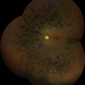
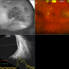
Fundus photograph of the left eye in a 72-year-old patient diagnosed with Stage I pancreatic carcinoma involving the head of the pancreas diagnosed three months ago. The patient was seen for a routine diabetic retinopathy evaluation. Incidentally, this mass was observed in the temporal periphery of the left eye. Visual acuity 20/25 OU. OCT normal OU.
Photographer: Amber Ozment
Imaging device: Optos California camera and Accutome 4 sight B scan ultrasonographer
Condition/keywords: choroidal metastasis
-
 Spontaneous Resolution of ERM
Dec 19 2020 by John S. King, MD
78-year-old diabetic with ERM OD and mild PCO OD that was stable for a few years, and eventually improved on own. Her acuity ranged from 20/25-2040 and she did not notice significant visual issues, and we decided to monitor. On her last visit she was 20/40 with 1+ PCO, and has not noticed any visual changes.
Imaging device: Zeiss Cirrus
Condition/keywords: epiretinal membrane (ERM), macular pucker
-
 MEWDS
MEWDS
Oct 9 2020 by David L Kilpatrick, MD
26-year-old female presented with unilateral vision loss. She c/o flashes and a peripheral scotoma. Vision was 20/100. On exam, she showed foveal granularity, mild disc edema, and white dots as seen. Three weeks later, white dots had resolved and vision improved to 20/25.
Photographer: Mississippi Retina Associates
Imaging device: Optos
Condition/keywords: multiple evanescent white dot syndrome (MEWDS)
-
 MEWDS - Widefield FA
Oct 9 2020 by David L Kilpatrick, MD
26-year-old female presented with unilateral vision loss. She c/o flashes and a peripheral scotoma. Vision was 20/100. On exam, she showed foveal granularity, mild disc edema, and white dots as seen. Three weeks later, white dots had resolved and vision improved to 20/25.
Photographer: Mississippi Retina Associates
Imaging device: Optos
Condition/keywords: multiple evanescent white dot syndrome (MEWDS)
 Loading…