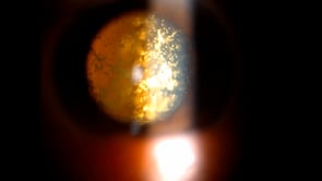
 Foreign Body in the Left Eye
Feb 9 2026 by Kimberly Wakester
Optomap RGB image of a 37-year-old man with a metallic foreign body in his left eye. The patient states the irritation and a new floater started after scraping ice off of his vehicle and something went flying into his eye. Patient was recommended to proceed with surgery to remove foreign body. After removal the foreign body was identified as a piece of steel. Patient is to continue post-operative care as instructed.
Photographer: Kimberly Wakester, COA, OCT-C
Imaging device: Optos California
Condition/keywords: metallic foreign body
 Proliferative Diabetic Retinopathy
Feb 9 2026 by Kimberly Wakester
Optomap FA image of a 69-year-old woman with proliferative diabetic retinopathy in the right eye. FA shows conversion to PDR with new areas of NVE inferonasal and capillary dropout. Treatment with PRP laser was recommended.
Photographer: Kimberly Wakester, COA, OCT-C
Imaging device: Optos California
Condition/keywords: FA, PDR, right eye
 Proliferative Diabetic Retinopathy
Feb 9 2026 by Kimberly Wakester
Optomap FA images of a 69-year-old woman with proliferative diabetic retinopathy in the right eye. The left image of the FA is captured at 37 seconds, the middle is captured at 2 minutes, and the right is captured at 5 minutes.
Photographer: Kimberly Wakester, COA, OCT-C
Imaging device: Optos California
Condition/keywords: FA early phase, FA late phase leakage, FA mid phase, PDR, right eye, type 2 diabetes with ocular complications
 Asteroid Hyalosis
Feb 9 2026 by Kimberly Wakester
Optomap RGB image of a 79-year-old man with asteroid hyalosis on the left eye. Observation recommended.
Photographer: Kimberly Wakester, COA, OCT-C
Imaging device: Optos California
Condition/keywords: Asteroid hyalosis, left eye
 Retinal Macroaneurysm
Feb 9 2026 by Kimberly Wakester
Optomap RGB image of a 73-year-old woman with a retinal macroaneurysm in her right eye, nasal to the optic nerve. Observation recommended and will return in 4 months to check for progression.
Photographer: Kimberly Wakester, COA, OCT-C
Imaging device: Optos California
Condition/keywords: retinal macroaneurysm, right eye
 Pigmentary Retinal Dystrophy
Feb 9 2026 by Kimberly Wakester
Optomap RGB montage photo of a 24-year-old woman with 360 Pigmentary Retinal Dystrophy. Unusual in appearance though differential could include congenital arrest of normal periphery, prior uveal effusion, peripheral dystrophy/atypical sectoral RP, etc. No treatment needed at this time, will monitor. Will recheck in 6 months.
Photographer: Kimberly Wakester, COA, OCT-C
Imaging device: Optos California
Condition/keywords: Pigmentary Retinal Dystrophy, right eye
 Pneumatic Retinopexy in Giant Retinal Tear with RD
Feb 8 2026 by Poornachandra B, MS, FVRS
A 24-year-old male presented with sudden onset distortion of vision in the right eye for one week. On evaluation, he was diagnosed with a giant retinal tear with associated retinal detachment in the right eye. He was managed with pneumatic retinopexy using 0.6 mL of SF6 gas. Following successful retinal reattachment, barrage laser photocoagulation was completed on day 2
Photographer: Mr Dhikshith
Condition/keywords: BARRAGE LASER, giant retinal tear, pneumatic retinopexy
 Serous Central Chorioretinopathy With Myelination of Retinal Nerve Fibers
Feb 6 2026 by Oftalmontt Clínica Láser
A 35 year-old patient showed a serous central chorioretinopathy with myelination of retinal nerve fibers in the right eye (temporal fiber raphe can be seen), together with myelination of nerve fibers of the left eye.
Photographer: Ophthalmic Medical Technologist
Imaging device: Canon cx-1
Condition/keywords: central serous chorioretinopathy (CSCR), mielinization
 Branch Retinal Vein Occlusion and Hypertensive Retinopathy
Feb 5 2026 by Kimberly Wakester
Optomap RGB and FA image of a 75-year-old woman with Branch retinal vein occlusion and Hypertensive retinopathy in the left eye. There is an area of significant partial venous occlusion with fine collateral vessels versus neovascularization along the STA in the left eye. On fluorescein angiogram there was no leakage seen from the telangiectatic vessels, however there is some vascular leakage super temporally. Since there is no edema or neovascularization present, observation was recommended. She is to return in 3-4 months for recheck, prn sooner if changes.
Photographer: Kimberly Wakester, COA, OCT-C
Imaging device: Optos California
Condition/keywords: BRVO, fluorescein angiogram (FA), hypertensive retinopathy, left eye, Optomap RGB
 Treated Horseshoe Tear of Retina Without Detachment
Feb 5 2026 by Kimberly Wakester
Optomap RGB image of an 55-year-old man with a treated horseshoe retinal tear in the right eye. The horseshoe tear remains well barricaded with laser; no extension of the tear past the laser on clinical exam and fundus photos. Observation recommended. Patient is to continue follow up care yearly.
Photographer: Kimberly Wakester, COA, OCT-C
Imaging device: Optos California
Condition/keywords: right eye, Treated Horseshoe Tear of Retina Without Detachment
 Retinal Detachment with Multiple Breaks
Feb 5 2026 by Kimberly Wakester
Optomap RGB image of a 58-year-old man with a retinal detachment with multiple breaks in the left eye. Patient presents with macula-involved RD extending from 3:00-8:00, tear from 4:00-5:00 within area of lattice degeneration. Surgery was recommended. Patient is to continue follow up care post operatively.
Photographer: Kimberly Wakester, COA, OCT-C
Imaging device: Optos California
Condition/keywords: left eye, macula-involved RD, Retinal Detachment with Multiple Breaks
 Chorioretinal Scar OD
Feb 5 2026 by Kimberly Wakester
Optomap RGB image of a 12-year-old boy with a Chorioretinal Scar in the right eye. Observation was recommended. Will continue follow up care yearly.
Photographer: Kimberly Wakester, COA, OCT-C
Imaging device: Optos California
Condition/keywords: chorioretinal scar, right eye
 Retinal Detachment With a Single Break
Feb 5 2026 by Kimberly Wakester
Optomap RGB image of a 64-year-old man with a retinal detachment with a single break in the right eye. Surgery was recommended. Patient is to continue follow up care post operatively.
Photographer: Kimberly Wakester, COA,
Imaging device: Optos California
Condition/keywords: Retinal Detachment, right eye
 Retinitis Pigmentosa
Feb 5 2026 by Kimberly Wakester
Optomap RGB and AF of a 72-year-old woman with Retinitis Pigmentosa. Patient is to continue follow up care every 12 months to monitor progression.
Photographer: Kimberly Wakester, COA, OCT-C
Imaging device: Optos California
Condition/keywords: autofluorescence imaging, Diffuse RPE atrophy, retinitis pigmentosa
 Intraocular Foreign Body with Hemorrhagic Choroidal Detachment
Feb 3 2026 by Poornachandra B, MS, FVRS
Optos image of a 25 year-old male with a history of ocular injury while chiselling metal, showing an intraocular foreign body in the inferotemporal quadrant with associated choroidal detachment
Photographer: Mr Dhikshith
Condition/keywords: choroidal detachment, intraocular foreign body
 Inferior Pediatric Retinal Detachment
Feb 2 2026 by Dallin Milner
12 year-old boy with chronic inferior retinal detachment with subretinal bands.
Condition/keywords: retinal detachment
 Sickle Cell Anemia
Feb 2 2026 by Adrienne W. Scott, MD, FASRS
Example images from right eye of a 13-year-old patient with HbSS. A. Areas of flow loss observed in the SCP and (B) DCP temporal to fovea on OCT-A (yellow arrows). Retinal temporal thinning is demonstrated (C) on cross-sectional OCT (yellow arrow) and (D) retinal thickness map (yellow arrow).
Condition/keywords: sickle cell
 Giant Retinal Tear
Feb 2 2026 by Cecilia Anggraini
Male, 39 years old, with rhegmatogenous retinal detachment with giant tear of the left eye.
Imaging device: Optos ultra wiled field
Condition/keywords: GIANT RETINAL TEAR
 Asteroid Hyalosis
Jan 30 2026 by Rim Kahloun Korbaa, MD
Asteroid hyalosis in the left eye of a 69-year-old male patient with a history of proliferative diabetic retinopathy treated with bilateral panretinal photocoagulation. The asteroid bodies exhibit synchronous movement with the vitreous and return to their original position following ocular movements. The vitreous is clear in the right eye.
Photographer: Rim Kahloun, MD
Condition/keywords: asteroid hyalosis, vitreous
 Subretinal Hemorrhage
Jan 30 2026 by Virginia Gebhart
63 year old male referred for expanding choroidal lesion. Large PED superior with sub-RPE oxidized hemorrhage and SRH. No vascularity on ultrasound, findings consistent with Peripheral Exudative Hemorrhagic Chorioretinopathy. Will observe.
Photographer: Virginia Gebhart, Retina Consultants of Carolina
Imaging device: Optos California
Condition/keywords: PED, peripheral exudative hemorrhagic chorioretinopathy (PEHCR), subretinal hemorrhage