Search results (43 results)
-
---thumb.jpg/image-square;max$300,300.ImageHandler) Adult Vitelliform Dystrophy
Adult Vitelliform Dystrophy
Mar 29 2013 by Henry J. Kaplan, MD
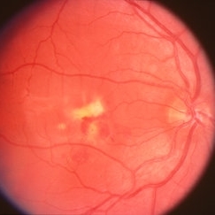
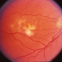
Fundus photograph of the same patient, left eye #2 notice the multiple vitelliform lesions.
Condition/keywords: vitelliform macular dystrophy
-
---thumb.jpg/image-square;max$300,300.ImageHandler) Adult Vitelliform Dystrophy
Adult Vitelliform Dystrophy
Apr 1 2013 by Henry J. Kaplan, MD
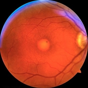
Fundus photograph of a middle aged patient with mild decreased vision and bilateral macular vitelliform lesion #1.
Condition/keywords: adult vitelliform dystrophy, vitelliform lesion, vitelliform macular dystrophy
-
---thumb.jpg/image-square;max$300,300.ImageHandler) Adult Vitelliform Dystrophy
Adult Vitelliform Dystrophy
Feb 13 2013 by From the Collections of Thomas M. Aaberg, MD and Thomas M. Aaberg Jr., MD
Right eye.
Condition/keywords: vitelliform macular dystrophy
-
---thumb.jpg/image-square;max$300,300.ImageHandler) Adult Vitelliform Dystrophy
Adult Vitelliform Dystrophy
Feb 13 2013 by From the Collections of Thomas M. Aaberg, MD and Thomas M. Aaberg Jr., MD
Left eye
Condition/keywords: left eye, vitelliform macular dystrophy
-
---thumb.jpg/image-square;max$300,300.ImageHandler) Adult Vitelliform Dystrophy
Adult Vitelliform Dystrophy
Feb 13 2013 by From the Collections of Thomas M. Aaberg, MD and Thomas M. Aaberg Jr., MD
Right eye.
Condition/keywords: vitelliform macular dystrophy
-
---thumb.jpg/image-square;max$300,300.ImageHandler) Adult Vitelliform Dystrophy
Adult Vitelliform Dystrophy
Feb 13 2013 by From the Collections of Thomas M. Aaberg, MD and Thomas M. Aaberg Jr., MD
Right eye.
Condition/keywords: vitelliform macular dystrophy
-
 Adult-onset foveomacular vitelliform dystrophy
May 26 2022 by Rinat Sutiushev
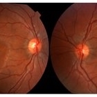
Woman born in 1946. Concerns about decreased vision in the right eye, distortions when reading. The ocular fundus of both eyes shows round yellowish deposits (vitelliform material deposits) in the fovea.
Photographer: Rinat Sutiushev
Condition/keywords: adult vitelliform dystrophy, vitelliform lesion, vitelliform macular dystrophy
-
 Adult-onset foveomacular vitelliform dystrophy
May 26 2022 by Rinat Sutiushev
Woman born in 1946. Concerns about decreased vision in right eye, distortions when reading. The ocular fundus of both eyes shows round yellowish deposits (vitelliform material deposits) in the fovea. When autofluorescence photography is performed, hyperautofluorescence is detected.
Photographer: Rinat Sutiushev
Condition/keywords: adult vitelliform dystrophy, vitelliform lesion, vitelliform macular dystrophy
-
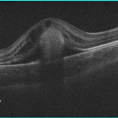
 Adult-onset foveomacular vitelliform dystrophy
May 26 2022 by Rinat Sutiushev
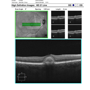
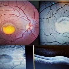
Patient born in 1946. Concerns about decreased vision in right eye, distortions when reading. The ocular fundus of both eyes shows round yellowish deposits (vitelliform material deposits) in the fovea. Autofluorescence photography reveals hyperautofluorescence. OCT demonstrates the presence of vitelliform material under the sensitive retina and over the retinal pigment epithelium.
Photographer: Rinat Sutiushev
Condition/keywords: adult vitelliform dystrophy, vitelliform lesion, vitelliform macular dystrophy
-
 Best Disease
Apr 8 2019 by Gary R. Cook, MD, FACS
Right eye of patient with Best disease with active CNV OD; V.A. = 20/30.
Condition/keywords: Best disease, choroidal neovascularization (CNV), vitelliform macular dystrophy
-
 Best Disease
Apr 8 2019 by Gary R. Cook, MD, FACS
Left eye of the patient with Best disease and an active CNV OD showing resolving hemorrhage from prior CNV OS.
Condition/keywords: Best disease, retinal hemorrhage, vitelliform macular dystrophy
-
 Best Disease
Jul 5 2014 by John S. King, MD
Late 30s.
Photographer: URMC
Condition/keywords: Best disease, vitelliform macular dystrophy
-
 Best Disease
Jul 5 2014 by John S. King, MD
30s.
Photographer: URMC
Condition/keywords: Best disease, vitelliform macular dystrophy
-
 Best Disease
Jul 5 2014 by John S. King, MD
20s.
Photographer: URMC
Condition/keywords: Best disease, vitelliform macular dystrophy
-
 Best Disease
Jul 5 2014 by John S. King, MD
Teens.
Photographer: URMC
Condition/keywords: Best disease, vitelliform macular dystrophy
-
 Best Disease
Oct 12 2012 by Gregg T. Kokame, MD, MMM, FASRS
Best disease
Photographer: Jaclyn Pisano, Retina Consultants of Hawaii
Imaging device: Zeiss FF-450 plus
Condition/keywords: Best disease, vitelliform macular dystrophy
-
 Best Disease
May 1 2018 by Mitzy E Torres Soriano, MD
Fundus photographs of an 5-year-old boy with best vitelliform macular dystrophy and family history.
Photographer: Luciana García,MD
Condition/keywords: Best disease, vitelliform macular dystrophy
-
 Best Vitelliform Dystrophy with Secondary CNVM
Jan 8 2019 by Rutul R Patel, MD Ophthalmology
10-year-old girl with b/l Best vitelliform dystrophy and left eye secondary CNVM.
Photographer: Dr. Rutul Patel
Imaging device: TOPCON TRC 50DX
Condition/keywords: Best disease, choroidal neovascular membrane (CNVM), vitelliform macular dystrophy
-
 Best Vitelliform Macular Dystrophy
Mar 17 2020 by Sophia El Hamichi, MD
Classic "egg yolk" presentation in a 16-year-old female with best disease.
Condition/keywords: autofluorescence imaging, Best disease, optical coherence tomography (OCT), vitelliform macular dystrophy
-
 Best Vitelliform Macular Dystrophy
Jan 14 2018 by Koushik Tripathy, MBBS, MD
Typical subretinal egg yolk appearance of Best disease. Arden ratio in electrooculogram was reduced in either eye.
Condition/keywords: Best disease, vitelliform macular dystrophy
-
 Best's Vitelliform Macular Dystrophy
Apr 8 2019 by Gary R. Cook, MD, FACS
Right eye of a 29-year-old white male with Best's disease; OD; VA = 20/25.
Condition/keywords: Best disease, vitelliform macular dystrophy
-
 Best's Vitelliform Macular Dystrophy
Apr 8 2019 by Gary R. Cook, MD, FACS
Left eye of 29-year-old white male with Best's disease showing only a little yellow material and more pigmentation of the lesion; VA = 20/200
Condition/keywords: Best disease, vitelliform macular dystrophy
-
 Best's Vitelliform Macular Dystrophy
Apr 8 2019 by Gary R. Cook, MD, FACS
Vitelliform lesion in the left eye of a 10-year-old female demonstrating a yellow, round, egg yolk-like lesion; V. A. = 20/25
Condition/keywords: Best disease, vitelliform lesion, vitelliform macular dystrophy
-
 Cystoid Macular Edema (CME) in Vitelliform Macular Dystrophy (VMD)
Apr 22 2018 by Ronald Silva
Macula OCT of a 3-year-old boy with low vision and cystoid macular edema (CME) in vitelliform macular dystrophy (VMD) in right eye.
Photographer: Ronald Rocha da Silva, HCOE, Feira de Santana-BA
Condition/keywords: Best disease, cystoid macular edema (CME), vitelliform macular dystrophy
-
 Vitelliform Macular Dystrophy
Sep 2 2012 by Hyung-Woo Kwak, MD
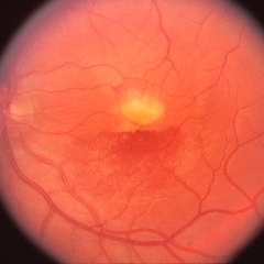
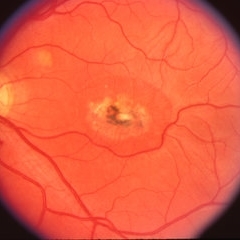
The typical appearance is of bilateral, round or oval, yellow, symmetrical, subretinal lesions, typically one-third to one-half optic disc diameter in size.
Imaging device: Zeiss F450 plus
 Loading…