Search results (75 results)
-
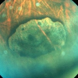
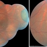
 CHRPE
CHRPE
Apr 1 2019 by Gary R. Cook, MD, FACS
35 year-old white female with asymptomatic CHRPE lesion OS; V.A.= 20/20-2.
Imaging device: Topcon VT-50
Condition/keywords: congenital hypertrophy of the retinal pigment epithelium (CHRPE)
-
 CHRPE
CHRPE
May 9 2014 by S. Natarajan, MD, FASRS, FRCS (GLASGOW) , FICO, D.Sc, FELA
23-year-old male in for routine eye checkup with BCVA 6/6 (OU). Showed CHRPE lesion in INQ (OD )on fundus.
Photographer: ADITYA JYOT EYE HOSPITAL,MUMBAI,INDIA
Condition/keywords: congenital hypertrophy of the retinal pigment epithelium (CHRPE), myopic eye
-
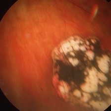
 CHRPE
CHRPE
Jun 3 2020 by stephen oconnell
Congenital hypertrophy of the RPE with minimal pigment
Condition/keywords: congenital hypertrophy of the retinal pigment epithelium (CHRPE)
-
 CHRPE
CHRPE
Jun 5 2020 by stephen oconnell
CHRPE
Condition/keywords: congenital hypertrophy of the retinal pigment epithelium (CHRPE)
-
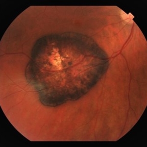
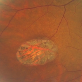
 CHRPE
CHRPE
Jan 12 2023 by Christopher R. Adam, M.D.
Optos color photograph of a 51 y/o F with a 9x9mm CHRPE in the nasal quadrant. The lesion is flat with a bordering halo and lacunae.
Condition/keywords: congenital hypertrophy of the retinal pigment epithelium (CHRPE)
-
 CHRPE
CHRPE
Dec 13 2018 by Jason Griffith
50-year-old male being followed for typical CHRPE presentation.
Photographer: Jason Griffith, Tennessee Retina, Nashville, TN
Imaging device: Clarus 500
Condition/keywords: congenital hypertrophy of the retinal pigment epithelium (CHRPE)
-
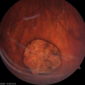
 CHRPE
CHRPE
Oct 8 2019 by DIEGO TOLENTINO
CHRPE plus laser barricade around retinal break
Photographer: Diego Tolentino
Condition/keywords: congenital hypertrophy of the retinal pigment epithelium (CHRPE)
-
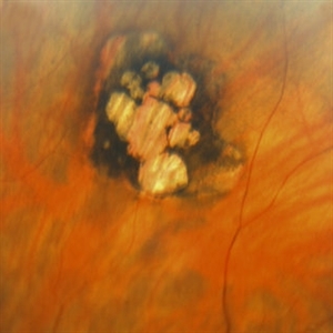
 CHRPE
CHRPE
Jan 15 2021 by Priya Rasipuram Chandrasekaran, MBBS, DO, DNB, FRCS
This is the fundus photo and fundus photo montage of the left eye of a 25-year-old male showing flat, solitary, round, greyish pigmented lesion situated AT THE equator with a scalloped margin. Vessels overlying the lesion are normal and there is a clear demarcation line between this and normal retina. The margins are hypopigmented with few hypopigmented lacunae inside.
Condition/keywords: congenital hypertrophy of the retinal pigment epithelium (CHRPE)
-
 CHRPE & Myelinated RNFL
May 21 2020 by John S. King, MD
47-year-old white female, asymptomatic, sent to evaluate a scar OD. 20/40 cc, normotensive, examination significant for a flat, solitary lesion with pigmented borders and depigmented center with early lacunae forming, along with myeliated RNFL at the temporal edge of the lesion.
Photographer: Kay Dalby
Imaging device: Topcon
Condition/keywords: congenital hypertrophy of the retinal pigment epithelium (CHRPE), myelinated nerve fiber layer
-
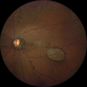
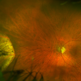
 Congenital Grouped Hypertrophy of RPE
Jun 7 2016 by Roy Schwartz, MD
54-year-old male with congenital grouped hypertrophy on routine examination.
Photographer: Galit Yair-Pur
Condition/keywords: congenital hypertrophy of the retinal pigment epithelium (CHRPE)
-
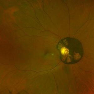
 Congenital Hypertrophy of RPE
Apr 2 2019 by Gary R. Cook, MD, FACS
67-year-old white male with typical CHRPE lesion demonstrating pigment hypertrophy and multiple lacunae in the inferotemporal periphery of OS. V.A. = 20/30
Imaging device: Topcon VT-50
Condition/keywords: congenital hypertrophy of the retinal pigment epithelium (CHRPE)
-
 Congenital Hypertrophy of RPE
Apr 2 2019 by Gary R. Cook, MD, FACS
67-year-old white male with small CHRPE lesion superiorly; V.A. = 20/25
Imaging device: Topcon VT-50
Condition/keywords: congenital hypertrophy of the retinal pigment epithelium (CHRPE)
-
 Congenital Hypertrophy of the Retinal Pigment (CHRPE)
Nov 21 2013 by Gavin Thorsrud
Congenital hypertrophy of the retinal pigment.
Photographer: Gavin Thorsrud, COMT, CRA
Imaging device: scanned slide - original image Topcon TRC 50 VT
Condition/keywords: congenital hypertrophy of the retinal pigment epithelium (CHRPE)
-
 Congenital Hypertrophy of the Retinal Pigment Epithelium (CHRPE)
Jul 14 2013 by Jason S. Calhoun
Fundus photo shows congenital hypertrophy of the retinal pigment epithelium (CHRPE), superior, nasally in the left eye.
Photographer: Jason S. Calhoun, Department of Ophthalmology, Mayo Clinic Jacksonville, Florida
Imaging device: TOPCON TRC 50-EX
Condition/keywords: congenital hypertrophy of the retinal pigment epithelium (CHRPE)
-
 Congenital hypertrophy of the retinal pigment epithelium (CHRPE)
Jun 22 2022 by Dawson Winter
Ultrawide field fundus autofluorescence optos image of the left eye of a 28 year old female. Patient admits to floaters that come and go, but has no other ocular symptoms at this time. At the time of the appointment the patient was seeing 20/30+1 OS. Patient underwent MRI testing of the ocular orbit and results were found to be normal.
Photographer: Dawson Winters
Imaging device: Optos California
Condition/keywords: autofluorescence imaging, choroidal lesions, congenital hypertrophy of the retinal pigment epithelium (CHRPE), fundus autofluorescence (FAF), left eye, Optos, temporal retina, ultra-wide field imaging
-
 Congenital Hypertrophy of the Retinal Pigment Epithelium (CHRPE)
Aug 24 2012 by Andrew N. Antoszyk, MD FASRS
CHRPE lesion (black pigmented lesion) located along superior temporal arcade of left eye
Photographer: Lorainne Clark, Charlotte Eye Ear Nose and Throat Associates
-
 Congenital Hypertrophy of the Retinal Pigment Epithelium (CHRPE)
Mar 1 2014 by Homayoun Tabandeh, MD, FASRS
Congenital hypertrophy of the retinal pigment epithelium (CHRPE).
Condition/keywords: congenital hypertrophy of the retinal pigment epithelium (CHRPE)
-
 Congenital Hypertrophy of the Retinal Pigment Epithelium (CHRPE)
Mar 1 2014 by Homayoun Tabandeh, MD, FASRS
Congenital hypertrophy of the retinal pigment epithelium (CHRPE).
Condition/keywords: congenital hypertrophy of the retinal pigment epithelium (CHRPE)
-
 Congenital Hypertrophy of the Retinal Pigment Epithelium Autofluorescence Optomap
Sep 24 2019 by Sophia El Hamichi, MD
A 52-year-old female followed for 2 temporal lesions of CHRPE OD and white without pressure.
Photographer: Sophia El Hamichi, MD, Murray Ocular Oncology and Retina, Miami
Condition/keywords: autofluorescence imaging, congenital hypertrophy of the retinal pigment epithelium (CHRPE), Optomap, ultra-wide field imaging, white without pressure
-
 Congenital Hypertrophy of the Retinal Pigment Epithelium Wide Field Optomap
Sep 24 2019 by Sophia El Hamichi, MD
A 52-year-old female followed for 2 temporal lesions of CHRPE OD and white without pressure.
Photographer: Sophia El Hamichi,MD, Murray Ocular Oncology and Retina, Miami
Condition/keywords: congenital hypertrophy of the retinal pigment epithelium (CHRPE), Optomap, ultra-wide field imaging, white without pressure
-
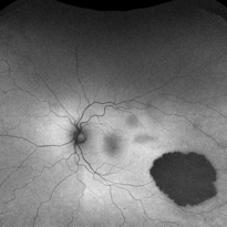
 Depigmented CHRPE
Nov 9 2022 by Maxwell J Wingelaar, MD
67 year-old female with a depigmented CHRPE
Condition/keywords: congenital hypertrophy of the retinal pigment epithelium (CHRPE)
-
---thumb.jpg/image-square;max$300,300.ImageHandler) Macular CHRPE
Macular CHRPE
Aug 11 2013 by Eric M. Shrier, DO
This a color fundus photograph of a 74-year-old black male with longstanding poor vision os, 20/200. He exhibits mild NPDR additionally.
Photographer: Christopher Bunce
Condition/keywords: congenital hypertrophy of the retinal pigment epithelium (CHRPE)
-
 Multimodal Imaging in CHRPE
Mar 6 2025 by Gerardo - Montante Montelongo, MD
Fundus photograph of an 83-year-old male with a history of Diabetes, smoking, cataract surgery on the right eye in 2022, and open-angle glaucoma. Asymptomatic. Indirect ophthalmoscopy revealed 80% excavation, peripapillary atrophy, and a hyperpigmented perifoveal lesion with 35% atrophy, 10% drusen, and 5.1 mm diameter, corresponding to a CHRPE. At multimodal imaging, FFA shows hypoautofluorescence of the lesion, OCT shows preservation of internal retinal layers, atrophy of external retinal layer, with an RPE disruption, and posterior shadowing. USG shows a flat hyperechoic lesion 5.1 mm in diameter and 1.32 mm in thickness, solid and with high internal reflectance.
Photographer: Gerardo Montante-Montelongo, MD, Mexican Institute of Ophthalmology
Imaging device: Clarus 700
Condition/keywords: congenital hypertrophy of the retinal pigment epithelium (CHRPE), multimodal imaging
-
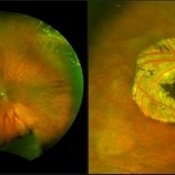 Operculated Hole and CHRPE
Jan 16 2018 by Carolyn Daley
58-year-old woman with an operculated hole and CHRPE in the right eye. Patient is asymptomatic so no treatment was recommended at this time.
Photographer: Carolyn Daley
Imaging device: Optos ultra wide field image
Condition/keywords: congenital hypertrophy of the retinal pigment epithelium (CHRPE), operculated retinal hole, Optos, ultra-wide field imaging
-
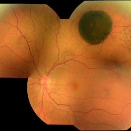
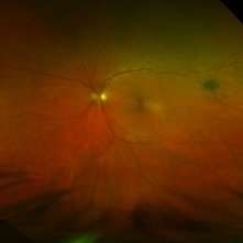
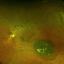
 Peripapillary Congenital Hypertrophy of the Retinal Pigment Epithelium
Dec 15 2022 by Jason Hsu, MD
Optos image of 62 year-old woman with incidental peripapillary congenital hypertrophy of the retinal pigment epithelium.
Photographer: Donnamarie Nielsen, COA
Imaging device: Optos California
Condition/keywords: congenital hypertrophy of the retinal pigment epithelium (CHRPE)
 Loading…