Search results (45 results)
-
 Serpigenous Choroidopathy in a 68-Year-Old Male
Feb 15 2013 by Roy Schwartz, MD
A 68-year-old healthy male presented with a few years of decreased vision bilaterally. Visual acuity in OD was 1/36 and in OS 20/40. Anterior segments were normal except for bilateral mild nuclear sclerosis and pseudoexfoliation in OS. In the fundus of OD a large atrophy with pigmentary scars were seen in the macula and nasally to the optic disc while OS presented with the same clinical picture but an island of normal appearing retina was seen in the fovea. On fluorscein angiography no leakage was shown. A diagnosis of Serpigenous choroidopathy was made.
Photographer: Galit Yair-Pur
Condition/keywords: macula serpiginous choroidopathy, serpiginous choroiditis
-
 Macular Serpiginous Choroidopathy
Sep 27 2012 by Raj K. Maturi, MD
10/21/2009
Photographer: Tom Steele, CRA
Imaging device: TRC 50ex
Condition/keywords: macula serpiginous choroidopathy
-
 Macular Serpiginous Choroidopathy
Sep 27 2012 by Raj K. Maturi, MD
9/11/2012
Photographer: Char Harris
Imaging device: HRA
Condition/keywords: red-free
-
 Serpigenous Choroidopathy in a 68-Year-Old Male
Feb 15 2013 by Roy Schwartz, MD
A 68-year-old healthy male presented with a few years of decreased vision bilaterally. Visual acuity in OD was 1/36 and in OS 20/40. Anterior segments were normal except for bilateral mild nuclear sclerosis and pseudoexfoliation in OS. In the fundus of OD a large atrophy with pigmentary scars were seen in the macula and nasally to the optic disc while OS presented with the same clinical picture but an island of normal appearing retina was seen in the fovea. On fluorscein angiography no leakage was shown. A diagnosis of Serpigenous choroidopathy was made.
Photographer: Galit Yair-Pur
Condition/keywords: macula serpiginous choroidopathy, serpiginous choroiditis
-
 Macular Serpiginous Choroidopathy
Sep 27 2012 by Raj K. Maturi, MD
10/21/2009
Photographer: Tom Steele, CRA
Imaging device: TRC 50ex
Condition/keywords: macula serpiginous choroidopathy
-
 Macular Serpiginous Choroidopathy
Sep 27 2012 by Raj K. Maturi, MD
10/21/2009
Photographer: Tom Steele, CRA
Imaging device: TRC 50ex
Condition/keywords: macula serpiginous choroidopathy
-
 Macular Serpiginous Choroidopathy
Sep 27 2012 by Raj K. Maturi, MD
9/11/2012
Photographer: Char Harris
Imaging device: HRA
Condition/keywords: serpiginous choroiditis
-
 Macular Serpiginous Choroidopathy
Sep 27 2012 by Raj K. Maturi, MD
9/11/2012
Photographer: Char Harris
Imaging device: HRA
Condition/keywords: macula serpiginous choroidopathy
-
 Macula Serpiginous Choroidopathy
Aug 27 2015 by Ruben A. Grigorian, MD
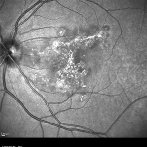
Red free photograph of an 18-year-old with macular serpiginous choroidopathy.
Photographer: Phylicia Yanna, Retina Eye Center, Eye Associates of Northeast Louisiana
Imaging device: Heidelberg Spectralis
Condition/keywords: macula serpiginous choroidopathy
-
 Macula Serpiginous Choroidopathy
Sep 27 2012 by Raj K. Maturi, MD
9/11/2012
Photographer: Char Harris
Imaging device: HRA
Condition/keywords: IR
-
---thumb.jpg/image-square;max$300,300.ImageHandler) Serpiginous Choroidopathy With Macular Scarring
Serpiginous Choroidopathy With Macular Scarring
Aug 1 2013 by From the Collections of Thomas M. Aaberg, MD and Thomas M. Aaberg Jr., MD
Serpiginous choroidopathy with macular scarring.
Condition/keywords: macular scar, serpiginous choroiditis
-
 Persistent Placoid Maculopathy (PPM)
Sep 5 2015 by Ali Tavallali, MD, FASRS
A 47-year-old female, with 20/200 VA of both eyes, stable for 23 years.
Photographer: Maryam Ravanshid
Condition/keywords: macula serpiginous choroidopathy
-
---thumb.jpg/image-square;max$300,300.ImageHandler) Serpiginous Choroidopathy With Macular Scarring
Serpiginous Choroidopathy With Macular Scarring
Aug 1 2013 by From the Collections of Thomas M. Aaberg, MD and Thomas M. Aaberg Jr., MD
Serpiginous choroidopathy with macular scarring.
Condition/keywords: macular scar, serpiginous choroiditis
-
 Macula Serpiginous Choroidopathy
Aug 27 2015 by Ruben A. Grigorian, MD
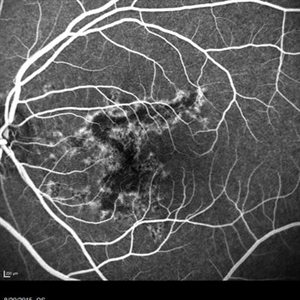
Fluorescein angiography of an 18-year-old with macular serpiginous choroidopathy.
Photographer: Phylicia Yanna, Retina Eye Center, Eye Associates of Northeast Louisiana
Imaging device: Heidelberg Spectralis
Condition/keywords: macula serpiginous choroidopathy
-
 Serpiginous Choroiditis (Recurrent)
Sep 22 2019 by Haider Ali
35-year-old female presented with decrease in vision in her left eye for last 4 days and in right eye for last 8 days. Her right eye was previously involved in a similar episode about 5-6 months ago for which she was treated with oral steroids.
Photographer: Dr Haider Ali Chaudhry, Madinah Teaching Hospital, Faisalabad
Condition/keywords: acute posterior multifocal placoid pigment epitheliopathy (APMPPE), macula serpiginous choroidopathy, serpiginous choroiditis, white dot syndrome
-
 Macular Serpiginous Choroidopathy
-
 Serpiginous Choroidal Atrophy
Mar 29 2019 by Jessica Norkus
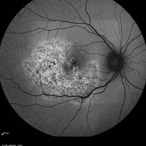
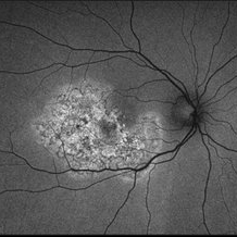
50 degree Auto fluorescent image of 20-year-old female presenting with serpiginous choroidal atrophy. Patient was unaware of vision loss OD, until accidentally covering OS and noticing the change. Acuity of 20/200 OD and 20/15 OS at time of imaging.
Photographer: Jessica Norkus
Imaging device: Heidelberg Spectralis
Condition/keywords: Heidelburg Spectralis, macula lesion, macula serpiginous choroidopathy, optical coherence tomography (OCT), wide angle imaging
-
 Serpiginous Choroiditis
Sep 22 2019 by Haider Ali
35-year-old female presented with decrease in vision in her left eye for last 4 days and in right eye for last 8 days. Her right eye was previously involved in a similar episode about 5-6 months ago for which she was treated with oral steroids.
Photographer: Dr Haider Ali Chaudhry, Madinah Teaching Hospital, Faisalabad
Condition/keywords: acute posterior multifocal placoid pigment epitheliopathy (APMPPE), macula serpiginous choroidopathy, posterior uveitis, serpiginous choroiditis, uveitis, white dot lesions, white dot syndrome
-
 Macular Serpiginous Choroidopathy
-
 Serpiginous Choroidal Atrophy
Mar 29 2019 by Jessica Norkus
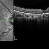
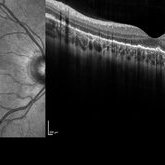
Heidelberg single horizontal scan image of 20-year-old female presenting with serpiginous choroidal atrophy. Patient was unaware of vision loss OD, until accidentally covering OS and noticing the change. Acuity of 20/200 OD and 20/15 OS at time of imaging.
Photographer: Jessica Norkus
Imaging device: Heidelberg Spectralis
Condition/keywords: Heidelburg Spectralis, macula lesion, macula serpiginous choroidopathy, optical coherence tomography (OCT)
-
 Serpiginous Choroidal Atrophy
Mar 29 2019 by Jessica Norkus
Optos ultra wide field auto fluorescent image of 20-year-old female presenting with serpiginous choroidal atrophy. Patient was unaware of vision loss OD, until accidentally covering OS and noticing the change. Acuity of 20/200 OD and 20/15 OS at time of imaging.
Photographer: Jessica Norkus
Imaging device: Optos Ultra Wide Field Camera
Condition/keywords: fundus autofluorescence (FAF), fundus photograph, macula lesion, macula serpiginous choroidopathy, Optos, ultra-wide field imaging
-
 Serpiginous Choroiditis
Sep 22 2019 by Haider Ali
35-year-old female presented with decrease in vision in her left eye for last 4 days and in right eye for last 8 days. Her right eye was previously involved in a similar episode about 5-6 months ago for which she was treated with oral steroids.
Photographer: Dr Haider Ali Chaudhry, Madinah Teaching Hospital, Faisalabad
Condition/keywords: acute posterior multifocal placoid pigment epitheliopathy (APMPPE), macula serpiginous choroidopathy, posterior uveitis, serpiginous choroiditis, uveitis, white dot lesions, white dot syndrome
-
 Serpiginous Choroidal Atrophy
Mar 29 2019 by Jessica Norkus
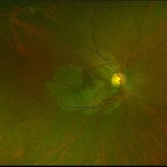
Optos ultra wide field color image of 20-year-old female presenting with serpiginous choroidal atrophy. Patient was unaware of vision loss OD, until accidentally covering OS and noticing the change. Acuity of 20/200 OD and 20/15 OS at time of imaging.
Photographer: Jessica Norkus
Imaging device: Optos Wide Field Camera
Condition/keywords: abnormal fundus, color fundus photograph, fundus photograph, macula serpiginous choroidopathy, Optomap, Optos, ultra-wide field imaging
-
 Serpiginous Choroidal Atrophy
Mar 29 2019 by Jessica Norkus
Heidelberg single vertical scan image of 20-year-old female presenting with serpiginous choroidal atrophy. Patient was unaware of vision loss OD, until accidentally covering OS and noticing the change. Acuity of 20/200 OD and 20/15 OS at time of imaging.
Photographer: Jessica Norkus
Imaging device: Heidelberg Spectralis
Condition/keywords: Heidelburg Spectralis, macula lesion, macula serpiginous choroidopathy, optical coherence tomography (OCT)
 Loading…