Search results (45 results)
-
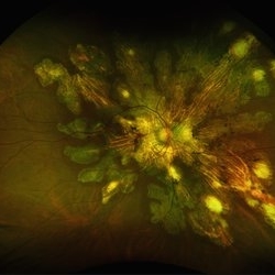
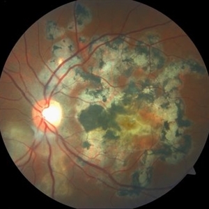
 Serpiginous Choroidopathy
Jun 23 2025 by César Adrián Gómez Valdivia, MD
Fundus photograph of a 29 year-old female patient diagnosed with Serpiginous Choroidopathy. Finings were bilateral. The most common complication of SC is choroidal neovascularization affecting up to 35% of patients. Other reported complications are subretinal fibrosis, cystoid macular edema, branch vein occlusion, serous retinal detachment, optic disc neovascularization ,and anterior uveitis.
Photographer: @eyemissu2
Imaging device: TOPCON TRC-50DX
Condition/keywords: serpiginous choroiditis
-
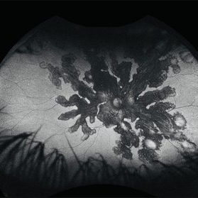
 Serpiginous Choroidopathy
Jun 23 2025 by César Adrián Gómez Valdivia, MD
Fundus photograph of a 29 year-old female patient diagnosed with Serpiginous Choroidopathy. Finings were bilateral. The most common complication of SC is choroidal neovascularization affecting up to 35% of patients. Other reported complications are subretinal fibrosis, cystoid macular edema, branch vein occlusion, serous retinal detachment, optic disc neovascularization, and anterior uveitis.
Photographer: @eyemissu2
Imaging device: California ICG OPTOS
Condition/keywords: serpiginous choroiditis
-
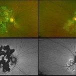
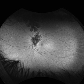 Serpiginous Choroidopathy
Jun 14 2025 by César Adrián Gómez Valdivia, MD
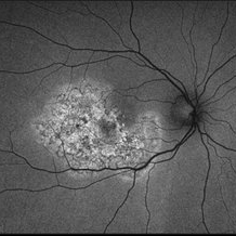
Fundus Autofluorescence of a 29-year-old woman with Serpiginous Choroidopathy. Finings were bilateral.
Photographer: @eyemissu2
Imaging device: California ICG OPTOS
Condition/keywords: Serpiginous Choroidopathy
-
 Serpiginous Choroidopathy
Jun 14 2025 by César Adrián Gómez Valdivia, MD
Fundus Autofluorescence of a 29-year-old woman with Serpiginous Choroidopathy. Finings were bilateral.
Photographer: @eyemissu2
Imaging device: California ICG OPTOS
Condition/keywords: Serpiginous Choroidopathy
-
 Serpiginous Choroidopathy
Oct 19 2024 by César Adrián Gómez Valdivia, MD
Fundus photograph of a 29-year-old woman with Serpiginous Choroidopathy. Finings were bilateral.
Photographer: @eyemissu2
Imaging device: California ICG OPTOS
Condition/keywords: macula serpiginous choroidopathy, serpiginous choroiditis, serpiginous like choroiditis
-
 Serpiginous Choroidopathy Autofluorescence
Sep 24 2024 by Gustavo Uriel Fonseca Aguirre
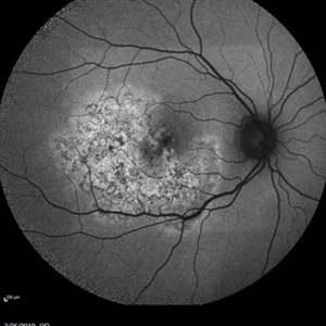
Autofluorescence image of the right fundus of a 32-year-old female patient diagnosed with serpiginous choroiditis.
Photographer: Gustavo U. Fonseca Aguirre, Fundación Hospital Nuestra Señora de la Luz, Ciudad de México
Condition/keywords: autofluorescence imaging, serpiginous choroiditis
-
 Serpiginous Choroidopathy
Sep 24 2024 by Gustavo Uriel Fonseca Aguirre
Right fundus of a 32-year-old female patient diagnosed with serpiginous choroiditis.
Photographer: Gustavo U. Fonseca Aguirre, Fundación Hospital Nuestra Señora de la Luz, Ciudad de México
Condition/keywords: Fundus examination, serpiginous choroiditis
-
 Serpiginous Choroidal Atrophy
May 28 2024 by Angela Rico
33 year-old female. Negative For TB or History of Immunosuppression. VA: OD 20/60-2 OS 20/150
Condition/keywords: macula serpiginous choroidopathy, serpiginous choroiditis
-
 Serpiginous Choroidal Atrophy
May 28 2024 by Angela Rico
33 year-old female. Negative For TB or History of Immunosuppression. VA: OD 20/60-2 OS 20/150
Photographer: Angela Rico M.D.
Condition/keywords: macula serpiginous choroidopathy, serpiginous choroiditis
-
 Serpiginous Choroidal Atrophy & CNV OS
May 15 2024 by Angela Rico
33 year-old female. Negative TB. No history of Immunosuppression.
Photographer: Angela Rico M.D.,
Condition/keywords: macula serpiginous choroidopathy, serpiginous choroiditis
-
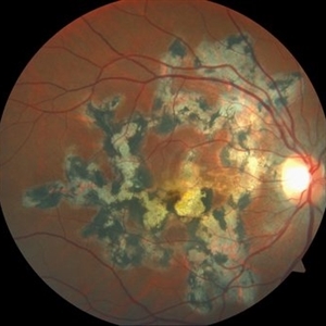
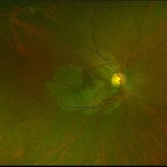
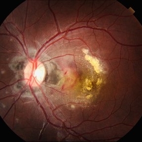 Serpiginous Choroidopathy
Apr 21 2024 by César Adrián Gómez Valdivia, MD
Gray-yellowish subretinal infiltrates that usually spread centrifugally from the peripapillary region in a serpiginous (snake-like) manner. Active lesions show a leading edge and resolve with subsequent RPE and choriocapillary atrophy.
Photographer: @eyemissu2
Imaging device: TOPCON TRC-50DX
Condition/keywords: macula serpiginous choroidopathy
-
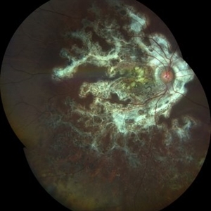 Serpiginous Choroidopathy
Mar 21 2024 by Ogugua Ndubuisi Okonkwo, MD, FRCS (Edin), FASRS
This is a right eye widefield fundus photograph of a 13-year-old male with a peripapillary ring of fibrotic scar that extends subretinally in finger-like projects along the vascular arcades and into the macula, with an extension of the scarring into the inferior retina, where it appears as a pigmented mottling.
Photographer: Zainab Ogunsanu, Eye Foundation Hospital & Eye Foundation Retina Institute, Lagos
Imaging device: ZEISS CLARUS 700
Condition/keywords: serpiginous like choroiditis
-
 Serpiginous Choroiditis (Recurrent)
Sep 22 2019 by Haider Ali
35-year-old female presented with decrease in vision in her left eye for last 4 days and in right eye for last 8 days. Her right eye was previously involved in a similar episode about 5-6 months ago for which she was treated with oral steroids.
Photographer: Dr Haider Ali Chaudhry, Madinah Teaching Hospital, Faisalabad
Condition/keywords: acute posterior multifocal placoid pigment epitheliopathy (APMPPE), macula serpiginous choroidopathy, serpiginous choroiditis, white dot syndrome
-
 Serpiginous Choroiditis
Sep 22 2019 by Haider Ali
35-year-old female presented with decrease in vision in her left eye for last 4 days and in right eye for last 8 days. Her right eye was previously involved in a similar episode about 5-6 months ago for which she was treated with oral steroids.
Photographer: Dr Haider Ali Chaudhry, Madinah Teaching Hospital, Faisalabad
Condition/keywords: acute posterior multifocal placoid pigment epitheliopathy (APMPPE), macula serpiginous choroidopathy, posterior uveitis, serpiginous choroiditis, uveitis, white dot lesions, white dot syndrome
-
 Serpiginous Choroidal Atrophy
Mar 29 2019 by Jessica Norkus
Optos ultra wide field auto fluorescent image of 20-year-old female presenting with serpiginous choroidal atrophy. Patient was unaware of vision loss OD, until accidentally covering OS and noticing the change. Acuity of 20/200 OD and 20/15 OS at time of imaging.
Photographer: Jessica Norkus
Imaging device: Optos Ultra Wide Field Camera
Condition/keywords: fundus autofluorescence (FAF), fundus photograph, macula lesion, macula serpiginous choroidopathy, Optos, ultra-wide field imaging
-
 Serpiginous Choroidal Atrophy
Mar 29 2019 by Jessica Norkus
50 degree Auto fluorescent image of 20-year-old female presenting with serpiginous choroidal atrophy. Patient was unaware of vision loss OD, until accidentally covering OS and noticing the change. Acuity of 20/200 OD and 20/15 OS at time of imaging.
Photographer: Jessica Norkus
Imaging device: Heidelberg Spectralis
Condition/keywords: Heidelburg Spectralis, macula lesion, macula serpiginous choroidopathy, optical coherence tomography (OCT), wide angle imaging
-
 Serpiginous Choroidal Atrophy
Mar 29 2019 by Jessica Norkus
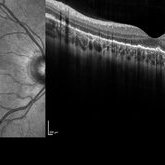
Heidelberg single vertical scan image of 20-year-old female presenting with serpiginous choroidal atrophy. Patient was unaware of vision loss OD, until accidentally covering OS and noticing the change. Acuity of 20/200 OD and 20/15 OS at time of imaging.
Photographer: Jessica Norkus
Imaging device: Heidelberg Spectralis
Condition/keywords: Heidelburg Spectralis, macula lesion, macula serpiginous choroidopathy, optical coherence tomography (OCT)
-
 Serpiginous Choroidal Atrophy
Mar 29 2019 by Jessica Norkus
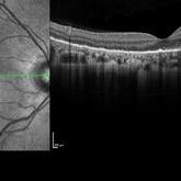
Heidelberg single horizontal scan image of 20-year-old female presenting with serpiginous choroidal atrophy. Patient was unaware of vision loss OD, until accidentally covering OS and noticing the change. Acuity of 20/200 OD and 20/15 OS at time of imaging.
Photographer: Jessica Norkus
Imaging device: Heidelberg Spectralis
Condition/keywords: Heidelburg Spectralis, macula lesion, macula serpiginous choroidopathy, optical coherence tomography (OCT)
-
 Serpiginous Choroidal Atrophy
Mar 29 2019 by Jessica Norkus
Optos ultra wide field color image of 20-year-old female presenting with serpiginous choroidal atrophy. Patient was unaware of vision loss OD, until accidentally covering OS and noticing the change. Acuity of 20/200 OD and 20/15 OS at time of imaging.
Photographer: Jessica Norkus
Imaging device: Optos Wide Field Camera
Condition/keywords: abnormal fundus, color fundus photograph, fundus photograph, macula serpiginous choroidopathy, Optomap, Optos, ultra-wide field imaging
-
 Persistent Placoid Maculopathy (PPM)
Sep 5 2015 by Ali Tavallali, MD, FASRS
A 47-year-old female, with 20/200 VA of both eyes, stable for 23 years.
Photographer: Maryam Ravanshid
Condition/keywords: macula serpiginous choroidopathy
-
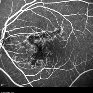 Macula Serpiginous Choroidopathy
Aug 27 2015 by Ruben A. Grigorian, MD
Fluorescein angiography of an 18-year-old with macular serpiginous choroidopathy.
Photographer: Phylicia Yanna, Retina Eye Center, Eye Associates of Northeast Louisiana
Imaging device: Heidelberg Spectralis
Condition/keywords: macula serpiginous choroidopathy
 Loading…