-
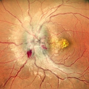
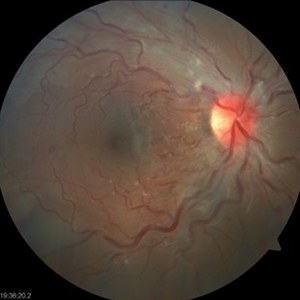
 Dropped Nucleus With Disc Pallor With Posterior Pole Retinal Detachment
Sep 12 2025 by Akansha Sharma
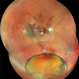
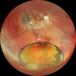
Color fundus photograph of a 60 year old male with a dropped nucleus with disc pallor with posterior pole retinal detachment.
Photographer: DR. AKANSHA SHARMA
Condition/keywords: dropped nucleus, fragmatome, nucleus drop, optic disc pallor, PALE DISC, POSTERIOR POLE RETINAL DETACHMENT, RD, retinal detachment
-
 Subhyaloid Hemorrhage With Vitreous Hemorrhage
Sep 12 2025 by Akansha Sharma
Color fundus photograph of a 56 year old hypertensive and diabetic female who presented with subhyaloid hemorrhage along with vitreous hemorrhage after being administered high dose anti-platelet therapy pre- and post a cardiac procedure.
Photographer: DR. AKANSHA SHARMA
Condition/keywords: SHH, sub ILM hemorrhage, subhyaloid hemorrhage, VH, vitreous hemorrhage
-
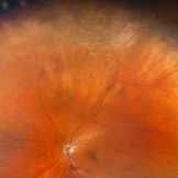
 Dislocated Nucleus
Sep 12 2025 by Tejaswita Verma
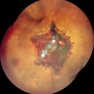
Fundus photo of a middle aged male with 6/36 vision, spontaneously dislocated nucleus posteriorly with focal retinal detachment. Right eye Pars plana Vitectomy + nucleus removal + intravitreal C3F8 (12%) gas was performed for this patient.
Photographer: DR. TEJASWITA VERMA
Imaging device: MIRANTE
Condition/keywords: dislocated lens, retinal detachment
-
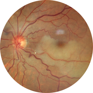
 Retinal Artery Macroaneurysm With Macular Edema
Sep 12 2025 by Tejaswita Verma
Fundus photo of a 73 year old hypertensive female with 6/18 vision, presenting with RAM ,with surrounding hard exudates and macular edema. She was advised focal laser, anti VEGF injection.
Photographer: DR. TEJASWITA VERMA
Imaging device: MIRANTE
Condition/keywords: RAM, retinal arterial macroaneurysm
-
 GRT Detachment of 10+ Clock Hours With Folded Retina
Sep 11 2025 by Luis J Haddock, MD
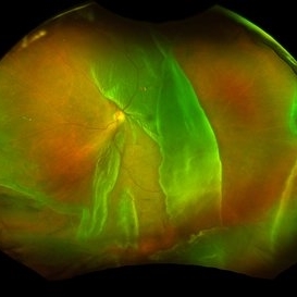
Fundus photo of giant retinal tear detachment involving 10+ hours of continuous tearing of the retina, visible anterior edge of retina over GRT.
Photographer: Natella Romero, University of Miami, Bascom Palmer Eye Institute
Imaging device: Optos
Condition/keywords: acute retinal detachment, Giant retinal tear
-
 Bilateral Disc Edema
Sep 11 2025 by rohan jain
bilateral disc edema
Photographer: Dr. ROHAN JAIN
Imaging device: mirante
Condition/keywords: disc edema
-
 Bilateral Disc Edema
Sep 11 2025 by rohan jain
bilateral disc edema
Photographer: Dr. ROHAN JAIN
Imaging device: mirante
Condition/keywords: disc edema
-
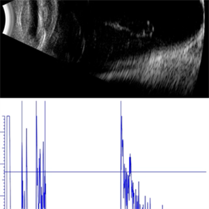 AL 39.16 mm
Sep 10 2025 by Gustavo Uriel Fonseca Aguirre
This axial B-scan reveals an elongated globe with an axial length of 39.16 mm, consistent with high axial myopia. Posterior staphyloma and scleral thinning are observed, though the retina remains attached.
Photographer: Gustavo U. Fonseca Aguirre, Hospital Conde de Valenciana, Ciudad de México
Condition/keywords: high myopia
-
 Recurrent Ciliary Body Melanoma After 19 Years
Sep 10 2025 by Virginia Gebhart
86 year old female with recurrence of ciliary body melanoma s/p sectoral iridectomy in 2006. Small pigmented lesion has shown unequivocal growth since initial presentation in 2024. Unclear if this is a new lesion or has been present for an extended time. Based on exam, diagnostics, and review of records, the determination was made for treatment by placement of radiation plaque for 4 days.
Photographer: Virginia Gebhart, Retina Consultants of Carolina
Imaging device: Ellex Eye Cubed
Condition/keywords: ciliary body melanoma, melanoma, recurrence, ultrasound biomicroscopy
-
 Optic Disc Neovascularization
Sep 9 2025 by Seif Allah Anwar
A case of high risk proliferative diabetic retinopathy with large disc neovascularization.
Photographer: Dr Seif Anwar
Imaging device: Topcon
Condition/keywords: optic disc neovascularization
-
 Large Leaking Optic Disc Neovascularization
Sep 9 2025 by Seif Allah Anwar
Large leaking optic disc neovascularization.
Photographer: Dr Seif Anwar
Imaging device: Topcon
Condition/keywords: Fundus Fluorescein Angiography
-
 Macular Tributary Retinal Venous Occlusion
Sep 7 2025 by Anand Temkar
A 55 yrs old female, k/c/o DM ( type II ) since past 5 yrs ( on medication ). Her vision was 6/6 in RE and 6/24 in her LE. IOP was 16 in both eyes. On examination, RE was WNL, and in LE ( color photo ) we noticed exudates, small hemorrhages, edema and sclerosed vessel ( depicted by black arrow. OCT LE shows altered foveal contour with cystic spaces and intraretinal hyperreflective material ( IRHRM ).
Photographer: Dr.Anand Temkar- Vasan Eye Hospital, Tiruchirappalli
Imaging device: Opticon
Condition/keywords: macular branch retinal vein occlusion (BRVO), venous occlusion
-
 Unexpected Sanctuary: Gas Bubble Entrapment in Morning Glory Disc
Sep 5 2025 by Danny Salgado Gómez
Fundus photograph of a 62-year-old male patient with Morning Glory syndrome in the right eye, who underwent vitrectomy, gas, and endolaser for posterior pole detachment. In the postoperative period, a gas bubble is observed within the optic disc, which persisted even after complete reabsorption of the intraocular gas.
Photographer: Dr. Danny Salgado, Retina and Vitreous Fellow, Clínica Oftalmológica del Caribe, Colombia.
Condition/keywords: gas bubble, intraocular gas, Morning Glory, Retinal Detachment, vitrectomy
-
 B-scan Ultrasound of Choroidal Melanoma with Serous Retinal Detachment
Sep 5 2025 by Kristen Wagner
B-scan ultrasound of a choriodal melanoma with serous retinal detachment.
Photographer: Kristen Wagner, COT Tennessee Retina
Condition/keywords: B scan ultrasound, Choroidal melanoma, serous retinal detachment
-
 Retinal Tear
Sep 4 2025 by Kimberly Wakester
Optomap RBG of a 55-year-old woman with a retinal tear at 12 with bridging vessel and some fluid. Treatment with prophylaxis laser was recommended. Patient is to continue follow up care post operatively.
Photographer: Kimberly Wakester, COA, OCT-C
Imaging device: Optos California
Condition/keywords: left eye, PVD, Retinal tear
-
 Fluorescein Angiography Papillophlebitis Salauno
Sep 3 2025 by Pablo Angel Garcia Uribe
In the arteriovenous phase, fluorescein angiography demonstrated venous engorgement and tortuosity, with relative incompetence of the venous walls leading to mild leakage. Optic disc staining with late leakage was also observed. There was no evidence of significant capillary non-perfusion, and only subtle perivenous leakage was noted. The foveal region remained spared.
Photographer: Optom. Marilyn Alvarez Monroy, Clínica Oftalmológica Salauno
Imaging device: Visucam 524, Carl Zeiss Meditec AG, Jena, Germany
Condition/keywords: FA late phase leakage, retina
-
 Papillophlebitis Salauno
Sep 3 2025 by Pablo Angel Garcia Uribe
Fundus photograph of a 24-year-old woman, previously healthy, with a history of recreational inhaled cannabis use, presented with a 24-hour history of photopsias and mild decrease in visual acuity, associated with a subtle relative central scotoma in the right eye. On ophthalmic examination, the anterior segment of both eyes was unremarkable. Best-corrected visual acuity was slightly reduced in the right eye and normal in the left. Fundus biomicroscopy of the right eye revealed moderate disc edema with hyperemia and well-defined margins, accompanied by venous engorgement and tortuosity, predominantly affecting the venules. No retinal hemorrhages were observed. Additionally, retinal thickening was noted along the temporal arcades, with apparent foveal sparing. The left eye showed no pathological findings. Based on the patient’s age, the acute onset of symptoms, the fundoscopic features, and the absence of systemic risk factors, the clinical presentation was consistent with papillophlebitis.
Photographer: Clínica Oftalmológica Salauno
Imaging device: Visucam 524, Carl Zeiss Meditec AG, Jena, Germany
Condition/keywords: papillophlebitis
-
 Hollenhorst Plaque
Sep 2 2025 by KANWALJEET HARJOT MADAN, M.S. (Ophthalmology); FAICO (Vitreous - Retina)
A 64 year-old male presented with sudden decrease in vision in LE for 1 week. His BCVA in LE was 20/200. Fundus exam revealed presence of whitish ischemic area in macula superior to fovea suggestive of branch retinal artery occlusion. A bright tiny refractile cholesterol embolus (Hollenhorst plaque) was visible in retinal artery. The patient was advised cardiology consultation.
Photographer: Dr. Kanwaljeet Harjot Madan, Thind Eye Hospital, Jalandhar City (Punjab). INDIA.
Imaging device: Zeiss Fundus Camera
Condition/keywords: branch retinal artery occlusion (BRAO), hollenhorst plaque
-
 Coats' Disease
Sep 2 2025 by Drew Mitchell
Optos color photograph of a young boy with Coats disease. Extensive subretinal exudation that is encroaching towards macula. There are peripheral berry aneurysms with localized area of subretinal fluid. Discussed treatment options including laser photocoagulation of aneurysms. Risks benefits and alternatives discussed including possible need for cryo.
Photographer: Drew Mitchell, OCT-C
Imaging device: Optos California
Condition/keywords: Coats' disease
-
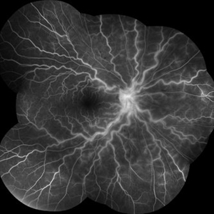
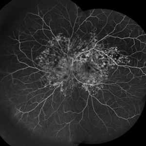 Snaking Away
Sep 1 2025 by Malvika Singh
Fluorescein angiography montage of a 45 year old man showing areas of staining in a case of healed choroiditis.
Photographer: Dr Malvika Singh, Retina Foundation, Ahmedabad, India
Imaging device: Mirante SLO/OCT
Condition/keywords: fluorescein angiogram (FA), healed choroiditis, serpiginous choroiditis
A project from the American Society of Retina Specialists
 Loading...