Search results (35 results)
-
 Dropped Crystalline Lens
Mar 8 2019 by Abdulaziz A. Alshamrani, MD
A 15-year-old female with congenital glaucoma complaining of acute diminution of vision after a blunt trauma.
Condition/keywords: crystalline lens, dropped nucleus, ora serrata
-
 Normal Nasal Ora Serrata
Nov 9 2012 by Norman Byer
This shows the normal nasal ora serrata. Note the dentate processes which divide the nasal ora into prominent bays and teeth
Condition/keywords: dentate processes, normal nasal ora serrata, ora bay, ora teeth
-
 Normal Temporal Ora Serrata
Nov 9 2012 by Norman Byer
This is the normal temporal ora serrata in a 26-year-old man. Note the typical ragged moth-eaten appearance caused by peripheral cystoid degeneration. This appearance may be present in infants but is always present beyond the age of eight years.
Condition/keywords: ora serrata, peripheral cystoid degeneration
-
 Congenital Meridional
Nov 9 2012 by Norman Byer
This is the same case as seen in the previous photograph but showing an area just below the lower end of the dialysis. It shows a congenital meridional fold at the 2 o’clock meridian with a retinal break at the posterior end possibly caused by the direct injury described previously.
Condition/keywords: meridional fold, ora serrata, retinal break
-
 From Ora to Ora
Aug 26 2024 by Nassim Alejandro Abreu Arbaje, MD
Ultra-wide field OCT angiography of a 39 year-old healthy male. The photo attempts to explore retinal vasculature up to the ora serrata.
Photographer: Johel Arrieta, TowardPi
Imaging device: TowardPi BMizar 400khz
Condition/keywords: OCT Angiography, OCTA, ultra-wide field imaging
-
 Normal Nasal Ora Serrata
Nov 9 2012 by Norman Byer
This is the normal nasal ora serrata showing a prominent meridional fold. Such folds are most commonly seen at the lower part of the upper nasal quadrant, and are present in 26% of the population. They are a normal developmental variation and are often bilateral.
Condition/keywords: meridional fold, normal developmental variation, normal nasal ora serrata, upper nasal quadrant
-
 Retinal Break
Nov 9 2012 by Norman Byer
This is the right eye of a 49-year-old woman showing a tiny retinal break adjacent to the temporal ora serrata. It has remained exactly the same without treatment for nine years.
Condition/keywords: ora serrata, retinal break
-
Time to Chill
Jan 23 2024 by SHISHIR VERGHESE, MS, FVRS, FAICO (Retina)
Intraoperative surgical video of a 65 year old female patient with advanced proliferative diabetic retinopathy showing neovascularization at the ora serrata for which a cryopexy is being done to cause regression. This video highlights a previously undocumented grape like Neovascularization at the ora serrata in this patient with advanced proliferative diabetic retinopathy.
Condition/keywords: Advanced Proliferative diabetic retinopathy, Cryopexy, neovascularization
-
 Cyst of the Pars Plana
Nov 9 2012 by Norman Byer
This is a cyst of the pars plana located just anterior to the ora serrata in the lower temporal quadrant. It illustrates how far anterior one may visualize the fundus with indirect ophthalmoscopy and scleral indentation. Pars plana cysts are common lesions of no particular clinical significance.
Condition/keywords: cyst of the pars plana, lower temporal quadrant, ora serrata, scleral indentation
-
 Pars Plana Cysts
Jan 29 2018 by Shani Pillar
During a pars plana vitrectomy for fixation of a dislocated IOL, this finding of pars plana cysts was seen, while performing indentation. Pars plana cysts are not uncommon, but rarely visualized so clearly, given their extremely peripheral location.
Photographer: Dr. Shani Pillar, Meir Medical Center, Kfar Saba, Israel
Imaging device: Intraoperative microscope
Condition/keywords: cyst of the pars plana, ora serrata, peripheral fundus lesion
-
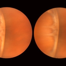
 A Classic Case of Retinal Ora Serrata Imaging
Jan 16 2025 by yuan duo
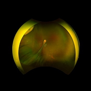
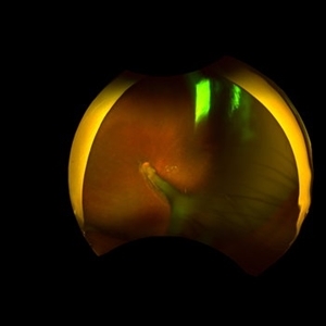
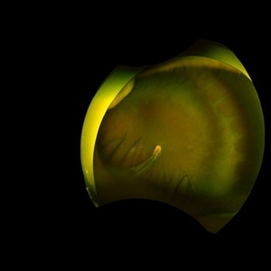
A 5-year-old girl, born full-term with no history of systemic disease, presented with poor vision since early childhood and underwent fundus examination. Anterior segments of both eyes showed no significant abnormalities. Fundus examination revealed retinal folds extending from the optic disc to the temporal peripheral retina, with blood vessels coursing through the folds (A, B). Avascular zones were observed in the peripheral retina, and the ora serrata’s boundaries were clearly visible, displaying dentate processes and bays (C, D). Retinal pigmentation was evident. Genetic testing confirmed the final diagnosis of bilateral Familial Exudative Vitreoretinopathy (FEVR).
Condition/keywords: Retinal Ora Serrata
-
 Complete PVD
Dec 10 2012 by Yale L. Fisher, MD
Dr. Yale Fisher presents a sagittal view of a complete posterior detachment demonstrated by the thin preretinal reflection (yellow arrow). Scleral depression (green arrow) at the ora serrata demonstrates the ability to register anatomical position on ultrasound using a scleral depressor.
Condition/keywords: video
-
 Dialysis of Retina in Upper Nasal Quadrant
Nov 9 2012 by Norman Byer
This 20-year-old wrestler sustained a sharp powerful blow to his right eye from his opponent’s thumb. One hour later he saw hundreds of black specs in his vision and was found to have this dialysis of his retina in the upper nasal quadrant. Note the triangular piece of retina in the center that remained attached to the ora serrata causing the retinal flap to resemble a man’s flared shirt collar.
Condition/keywords: retinal dialysis, retinal tear, upper nasal quadrant
-
 Elevated Cystic Area
Nov 9 2012 by Norman Byer
This is the eye of a 53-year-old woman with a small elevated cystic area of the peripheral retina at the posterior end of a meridional fold.
Condition/keywords: meridional fold, ora serrata, peripheral retina, small elevated cystic area
-
 Enclosed Ora Bay On The Temporal Side
Nov 9 2012 by Norman Byer
This is a developmental abnormality in a 59-year-old man. It is an enclosed ora bay on the temporal side, an isolated island of normal pars plana epithelium. It is important not to confuse this entity with a retinal break. It has smooth, sloping borders not a sharp, thin, visible retinal edge as a retinal break would have. The border looks exactly like that of the ora serrata, and the grayish pigmented base has the same appearance as the normal pars plana.
Condition/keywords: developmental abnormality, enclosed ora bay, grayish pigmented base, horizontal nasal meridian, pars plana epithelium, smooth sloping borders, temporal retina
-
 Enclosed Ora Bay On The Temporal Side
Nov 9 2012 by Norman Byer
This is another example of an enclosed ora bay on the temporal side. It is surrounded by normal retina and well separated from the ora serrata, which is toward the upper right just beyond the photograph. The yellow nubbin marks an abortive dentate process.
Condition/keywords: abortive dentate process, enclosed ora bay, normal eye, normal retina, ora serrata, temporal retina
-
 Enucleated Eye with Cataractous Lens
May 18 2020 by McGill University Health Centre
The cornea is transparent and thin. The lens is cataractous. The ora Serrata (arrow) demarcates a transition zone in the uveal tract between the pars plana of the ciliary body and the retina.
Condition/keywords: cataract, enucleation
-
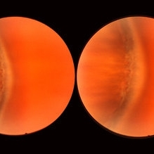
 Familial Exudative Vitreoretinopathy
Jan 16 2025 by yuan duo
A 5-year-old girl, born full-term with no history of systemic disease, presented with poor vision since early childhood and underwent fundus examination. Anterior segments of both eyes showed no significant abnormalities. Fundus examination revealed retinal folds extending from the optic disc to the temporal peripheral retina, with blood vessels coursing through the folds (A, B). Avascular zones were observed in the peripheral retina, and the ora serrata’s boundaries were clearly visible, displaying dentate processes and bays (C, D). Retinal pigmentation was evident. Genetic testing confirmed the final diagnosis of bilateral Familial Exudative Vitreoretinopathy (FEVR).
Condition/keywords: Retinal Ora Serrata
-
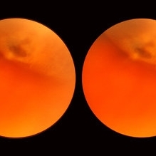
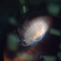
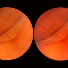
 Human Vitreous Base Structure
Sep 1 2020 by J. Sebag, MD, FACS, FRCOphth, FARVO
Dark-field slit microscopy was performed on fresh, unfixed, post-mortem human eyes that had undergone dissection to peel off the sclera, choroid, and retina. The vitreous body remains attached to the anterior segment which is seen below, while the posterior pole is above in these images. Left: specimen was tilted to reveal the posterior aspect of the lens (L) and the fibers of the vitreous base (arrow) splayed out to insert anterior and posterior to the ora serrata; Right: Anterior Loop of the vitreous base (see text). [From Sebag J: The Vitreous - Structure, Function, and Pathobiology. Springer-Verlag, New York, 1989, pp. 41 & 42; images © Springer Nature, reprinted with permission]
Condition/keywords: vitreous
-
 Intermediate Uveitis
May 18 2020 by McGill University Health Centre
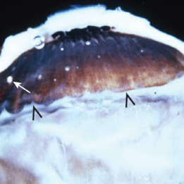
This enucleation specimen shows: “snowballs” or localized inflammatory foci (arrow); and a “snow bank” or inflammation at the ora serrata, the anterior-most limit of the retina. These are caused by a reaction to the subjacent uveitis (arrowhead).
Condition/keywords: intermediate uveitis
-
 Lattice Degeneration
Nov 9 2012 by Norman Byer
Lesion immediately adjacent to the ora serrata in an 18-year-old boy probably represents lattice degeneration characterized primarily by a reddish crater. It has remained unchanged for more than three years.
Condition/keywords: lattice degeneration, ora serrata, reddish crater
-
 Meridional Fold
Nov 9 2012 by Norman Byer
The next two photographs are of the same lesion in a 28-year-old woman. This view shows a sloping retinal mound with a radial retinal fold in the center. This is not a typical meridional fold for it stops short of the ora serrata and there is no dentate process. The upper temporal ora serrata and pars plana are well shown and peripheral cystoid degeneration is present posterior to the ora.
Condition/keywords: ora serrata, pars plana, peripheral cystoid degeneration, radial retinal fold, sloping retinal mound
-
 Pars Planitis - Peripheral Uveitis
Nov 9 2012 by Norman Byer
This 25-year-old man had pars planitis, peripheral uveitis bilaterally. In this eye it produced a small tractional oval tear of the retina and an inferior retinal detachment. The typical creamy yellow exudates of pars planitis can be seen in the lower right very close to the ora serrata.
Condition/keywords: creamy yellow exudates, inferior retinal detachment, pars planitis, peripheral uveitis, tractional retinal tear
 Loading…