A 57 year old man presented with painless progressive blurring of central vision OD of several years duration. He denied any history of hypertension or diabetes. Best corrected visual acuity was 20/200 OD and 20/20 OS.
He was noted to have telangectasia temporal to the fovea OD with unremarkable OS. OCT showed macular edema OD and was unremarkable OS. Fluorescein angiography confirmed telangectasia temporal to the fovea OD only with late leakage, consistent with macular edema. He underwent a series of antiVEGF injections, as well as focal laser. The macular edema resolved and the visual acuity improved from 20/200 to 20/20.
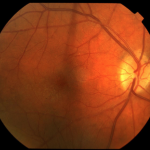
Idiopathic juxtafoveolar retinal telangiectasis was first described by Gass. Type 1 or unilateral juxtafoveal telangectasia typically affects men with a mean age of onset of 40 years. The telangiectasis occurs unilaterally and temporal to the fovea. Visual loss is mainly caused by macular edema and exudation.
-
 Idiopathic Juxtafoveal Telangectasia Type 1
Nov 4 2019 by Thomas A. Ciulla, MD, MBA, FASRS
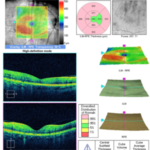
The telangiectasis occurs unilaterally in the temporal half of the macula in an area of 1–2 disc diameters. OCT originally showed significant macular edema temporally, mostly in the inner retina. He underwent a series of antiVEGF injections, as well as focal laser. The macular edema resolved and the visual acuity improved from 20/200 to 20/20.
Condition/keywords: idiopathic macular telangiectasia, juxtafoveal telangiectasis, parafoveal telangiectasia
-
 Idiopathic Juxtafoveal Telangectasia Type 1
Nov 4 2019 by Thomas A. Ciulla, MD, MBA, FASRS
The telangiectasis occurs unilaterally in the temporal half of the macula in an area of 1–2 disc diameters. OCT originally showed significant macular edema temporally, mostly in the inner retina. He underwent a series of antiVEGF injections, as well as focal laser. The macular edema resolved and the visual acuity improved from 20/200 to 20/20.
Condition/keywords: idiopathic macular telangiectasia, juxtafoveal telangiectasis, parafoveal telangiectasia
-
 Idiopathic Juxtafoveal Telangectasia Type 1
Oct 20 2015 by Thomas A. Ciulla, MD, MBA, FASRS
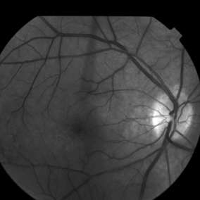
The telangiectasis occurs unilaterally in the temporal half of the macula in an area of 1–2 disc diameters. Vascular anomalies are noted on this red free image.
Photographer: Charlotte Harris
Condition/keywords: idiopathic macular telangiectasia, juxtafoveal telangiectasis, parafoveal telangiectasia
-
 Idiopathic Juxtafoveal Telangectasia Type 1
Oct 20 2015 by Thomas A. Ciulla, MD, MBA, FASRS
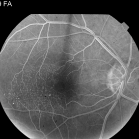
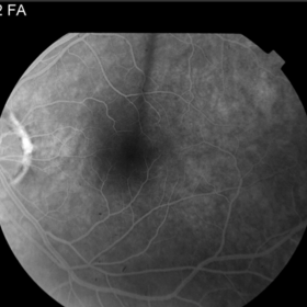
The telangiectasis occurs unilaterally in the temporal half of the macula in an area of 1–2 disc diameters. The anomalies are note in this early frame of the angiogram.
Photographer: Charlotte Harris
Condition/keywords: idiopathic macular telangiectasia, juxtafoveal telangiectasis, parafoveal telangiectasia
-
 Idiopathic Juxtafoveal Telangectasia Type 1
Oct 20 2015 by Thomas A. Ciulla, MD, MBA, FASRS
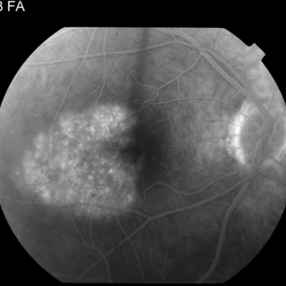
The telangiectasis occurs unilaterally in the temporal half of the macula in an area of 1–2 disc diameters. The anomalies begin to leak in this mid frame of the angiogram.
Photographer: Charlotte Harris
Condition/keywords: idiopathic macular telangiectasia, juxtafoveal telangiectasis, parafoveal telangiectasia
-
 Idiopathic Juxtafoveal Telangectasia Type 1
Oct 20 2015 by Thomas A. Ciulla, MD, MBA, FASRS
The telangiectasis occurs unilaterally in the temporal half of the macula in an area of 1–2 disc diameters. The late phase of the angiogram shows further leakage temporal to the fovea. Visual loss is mainly caused by macular edema and exudation.
Photographer: Charlotte Harris
Condition/keywords: idiopathic macular telangiectasia, juxtafoveal telangiectasis, parafoveal telangiectasia
-
 Idiopathic Juxtafoveal Telangectasia Type 1
Oct 20 2015 by Thomas A. Ciulla, MD, MBA, FASRS
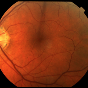
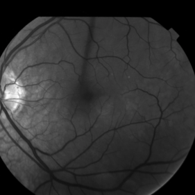
The fellow eye was unremarkable on this red free image.
Photographer: Charlotte Harris
Condition/keywords: idiopathic macular telangiectasia, juxtafoveal telangiectasis, parafoveal telangiectasia
-
 Idiopathic Juxtafoveal Telangectasia Type 1
Oct 20 2015 by Thomas A. Ciulla, MD, MBA, FASRS
The fellow eye was unremarkable on fluorescein angiography.
Photographer: Charlotte Harris
Condition/keywords: idiopathic macular telangiectasia, juxtafoveal telangiectasis, parafoveal telangiectasia
-
 Idiopathic Juxtafoveal Telangectasia Type 1
Nov 4 2019 by Thomas A. Ciulla, MD, MBA, FASRS
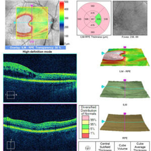
The telangiectasis occurs unilaterally in the temporal half of the macula in an area of 1–2 disc diameters. OCT shows macular edema temporally, mostly in the inner retina.
Condition/keywords: idiopathic macular telangiectasia, juxtafoveal telangiectasis, parafoveal telangiectasia
-
 Idiopathic Juxtafoveal Telangectasia Type 1
Nov 4 2019 by Thomas A. Ciulla, MD, MBA, FASRS
The telangiectasis occurs unilaterally in the temporal half of the macula in an area of 1–2 disc diameters. OCT originally showed significant macular edema temporally, mostly in the inner retina. He underwent a series of antiVEGF injections, as well as focal laser. The macular edema resolved and the visual acuity improved from 20/200 to 20/20.
Condition/keywords: juxtafoveal telangiectasis, parafoveal telangiectasia