-
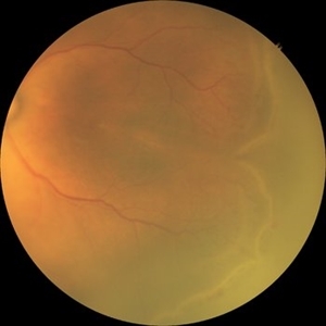
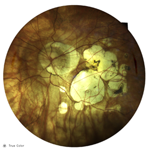
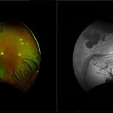
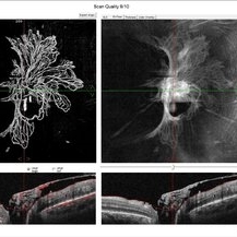 Peripapillary Neovessels
Oct 27 2025 by Oftalmontt Clínica Láser
OCT-A of a 54-year-old male patient with Proliferative Diabetic Retinopathy, observed the large number of neovessels in the optic disc along with fibrovascular proliferation.
Photographer: Ophthalmic Medical Technologist
Imaging device: Avanti XR AngioVue OptoVue
Condition/keywords: diabetic retinopathy
-
 Retinopathy of Prematurity
Oct 27 2025 by Anjana Mirajkar, MS Ophthalmology
Fundus photograph of a premature baby showing flat neovascularization with looping of the vessels with bleed in zone 1/2 with plus disease suggestive of A-ROP.
Photographer: Dr. Anjana Mirajkar- HV desai eye hospital ,Pune
Imaging device: Retcam
Condition/keywords: aggressive posterior retinopathy of prematurity (APROP)
-
 Retinopathy of Prematurity
Oct 26 2025 by Anjana Mirajkar, MS Ophthalmology
Fundus photograph of right eye of premature baby showing stage 3 in zone 2 posterior.
Photographer: Dr. Anjana Mirajkar- HV desai eye hospital ,Pune
Imaging device: Retcam
Condition/keywords: retinopathy of prematurity (ROP), stage 3
-
 Retinopathy of Prematurity
Oct 26 2025 by Anjana Mirajkar, MS Ophthalmology
Fundus photograph of a left eye of a premature baby showing stage 3 in zone 2 posterior.
Photographer: Dr. Anjana Mirajkar- HV desai eye hospital ,Pune
Imaging device: Retcam
Condition/keywords: retinopathy of prematurity (ROP), retinopathy of prematurity stage 3
-
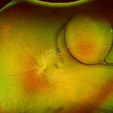
 Retinopathy of Prematurity
Oct 26 2025 by Anjana Mirajkar, MS Ophthalmology
Fundus photograph of left eye premature baby having stage 3 in zone 2A with a secondary notch.
Photographer: Dr. Anjana Mirajkar- HV Desai eye hospital ,Pune
Imaging device: retcam
Condition/keywords: retinopathy of prematurity (ROP), stage 3
-
 Posterior Dislocated Intraocular Lens
Oct 23 2025 by Aditya S Kelkar, MS, FRCS, FASRS,FRCOphth
Fundus photograph of an 53-year-old man with a posteriorly dislocated intraocular lens near the posterior pole.
Photographer: Dr Tejal Rao, National Institute of Ophthalmology, Pune, India
Imaging device: Optos Daytona
Condition/keywords: dislocated intraocular lens (IOL), IOL drop
-
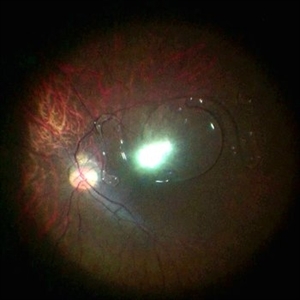 Dislocated ACIOL
Oct 23 2025 by KANWALJEET HARJOT MADAN, M.S. (Ophthalmology); FAICO (Vitreous - Retina)
This is intraoperative image of a young male who presented with sudden diminution of vision in RE after Trauma. Fundus exam revealed presence of dislocated anterior chamber IOL in Vitreous Cavity.
Photographer: Dr. Kanwaljeet Harjot Madan, Thind Eye Hospital, Jalandhar City (Punjab). INDIA.
Imaging device: Zeiss Fundus Camera
Condition/keywords: dislocated anterior chamber intraocular lens (ACIOL)
-
 Superior Temporal Venous Branch Occlusion
Oct 23 2025 by Vicente Nicanor Mancilla Guerrero
Confocal laser retinography of the right eye of a 45-year-old female patient with hypertension (+). Venous dilation is evidenced together with the presence of flame hemorrhages and cottony spots in the upper temporal arch.
Photographer: Vicente Mancilla G, Medical Technologist in Ophthalmology
Imaging device: Compass CenterVue
Condition/keywords: branch retinal vein occlusion (BRVO)
-
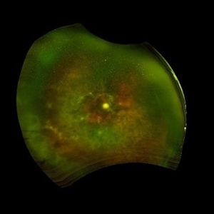
 Myopic Degeneration
Oct 23 2025 by Vicente Nicanor Mancilla Guerrero
Confocal laser retinography of a female patient with -20.00Dp myopia. Typical findings of myopic retinopathy can be seen.
Photographer: Vicente mancilla G, Ophthalmic Medical Technologist
Imaging device: Compass Centervue
Condition/keywords: high myopia
-
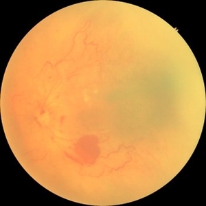
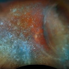 Choroidal and Near Total RD, Severe Asteroid Hyalosis, Treated Melanoma
Oct 22 2025 by Virginia Gebhart
78 year old male with sudden decrease in vision. Poor view due significant asteroid hyalosis. Bscan showed large nasal choroidal and near total retinal detachments, attached temporally. No obvious break found. Regressed tumor inferiorly s/p brachytherapy in April 2023. BCVA 20/320, IOP of 03. Pt schedule for primary PPV and possible SB placement vs. GFE
Photographer: Virginia Gebhart, Retina Consultants of Carolina
Imaging device: Optos California
Condition/keywords: asteroid hyalosis, brachytherapy, choroidal detachment, choroidal melanoma, melanoma, RD, retinal detachment, sub-total retinal detachment
-
 Proliferative Diabetic Retinopathy
Oct 22 2025 by Jeffrey Barker
56 year old Female with Diabetes Mellitus, lost to follow up for a year.
Photographer: Jeffrey P. Barker, B.S.
Condition/keywords: DME, fluorescein angiogram (FA), PDR
-
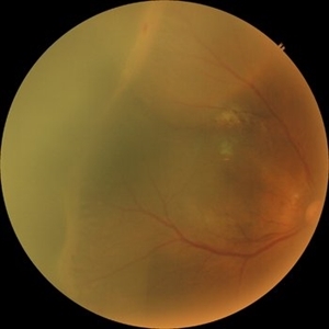
 Radiation Retinopathy
Oct 20 2025 by Meng-Hsin Chen
Fundus photograph of a 54-year-old woman with radiation retinopathy due to radiation exposure in-utero. Patient also presents with no light perception in the eye.
Photographer: Meng-Hsin Chen
Condition/keywords: No light perception, radiation retinopathy
-
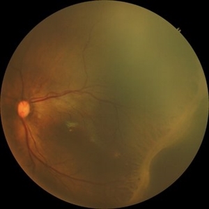
 Radiation Retinopathy with Rhegmatogenous Retinal Detachment
Oct 20 2025 by Meng-Hsin Chen
Fundus photo of a 54-year-old woman showing chronic radiation retinopathy from in-utero exposure with rhegmatogenous retinal detachment at the 3:00 -9:00 region, atrophic retina, and PVR. Lattice degeneration is present superiorly and inferiorly with a neovascular frond.
Photographer: Meng-Hsin Chen
Condition/keywords: atrophic retina, lattice degeneration, neovascular frond, proliferative vitreoretinopathy (PVR), radiation retinopathy, retinal detachment
-
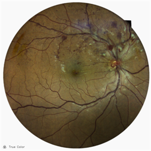
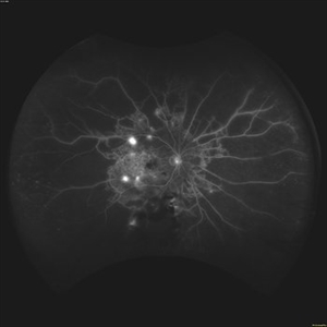
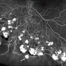
 Retinal Capillary Hemangioma
Oct 20 2025 by Jason Gayoski
Mid Stage FA of inferior retinal capillary hemangioma of the left eye.
Photographer: Jason Gayoski COA
Imaging device: Optos
Condition/keywords: retinal capillary hemangioma
-
 Retinal Capillary Hemangioma
Oct 20 2025 by Jason Gayoski
Late Stage FA of inferior retinal capillary hemangioma of the left eye.
Photographer: Jason Gayoski COA
Imaging device: Optos
Condition/keywords: retinal capillary hemangioma
-
 Retinal Capillary Hemangioma
Oct 20 2025 by Jason Gayoski
Early Stage FA of inferior retinal capillary hemangioma of the left eye.
Photographer: Jason Gayoski COA
Imaging device: Optos
Condition/keywords: retinal capillary hemangioma
-
 Retinal Capillary Hemangioma
Oct 20 2025 by Jason Gayoski
AF image of inferior retinal capillary hemangioma of the left eye
Photographer: Jason Gayoski COA
Imaging device: Optos
Condition/keywords: retinal capillary hemangioma
-
 Retinal Capillary Hemangioma
Oct 20 2025 by Jason Gayoski
Fundus image of inferior retinal capillary hemangioma of the left eye
Photographer: Jason Gayoski COA
Imaging device: Optos
Condition/keywords: retinal capillary hemangioma
-
 Retinal Capillary Hemangioma
Oct 20 2025 by Jason Gayoski
Transit Stage FA of inferior retinal capillary hemangioma of the left eye
Photographer: Jason Gayoski COA
Imaging device: Optos
Condition/keywords: retinal capillary hemangioma
-
 MEWDS
MEWDS
Oct 17 2025 by Jason Gayoski
32 year old female presenting to clinic with four day history of sudden onset unilateral left eye vision decrease with central scotoma upon awakening. VA OS 20/200 upon initial evaluation with wreath-like pattern of white dots surrounding macula OS. OD unaffected and asymptomatic.
Photographer: Jason Gayoski COA, Geisinger Ophthalmology
Imaging device: Heidelberg Spectralis
Condition/keywords: multiple evanescent white dot syndrome (MEWDS)
A project from the American Society of Retina Specialists
 Loading...